How is the diagnosis of myocarditis made?
introduction
Since myocarditis is a serious disease to be taken seriously, it is very important that if there is any suspicion, a thorough diagnosis is made and the myocarditis is not overlooked. The diagnosis of myocarditis is determined by one of the following:
- anamnese
- physical examination
- Blood test
- EKG
- Imaging procedures (ultrasound, MRI, X-ray)
The points are discussed in more detail below.
You might also be interested in this topic:
- Myocarditis
- Symptoms of myocarditis
- Myocarditis from exercise

anamnese
As with any disease, the diagnosis begins with the anamnesis. The symptoms of myocarditis are very variable.
This can be indicated by:
- fatigue
- Drop in performance
- Racing heart
- Chest pain
- Shortness of breath
The inflammation of the heart muscle can mimic heart failure. Their symptoms include shortness of breath and a drop in performance:
- Edema especially in the legs
- pulmonary edema is possible
- Cardiac arrhythmias
- increased need to urinate at night.
The symptoms often appear a few days to weeks after an upper respiratory tract infection.
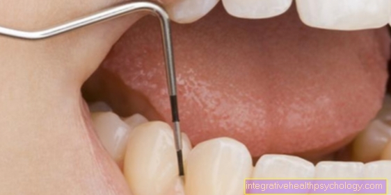
If the anamnesis is indicative for a heart muscle inflammation, it must be investigated further. As a rule, following the anamnesis, a physical examination and auscultation of the heart with the help of a stethoscope are carried out before further diagnostic equipment is carried out.
Read more on this topic at: Symptoms of myocarditis
Physical examination and auscultation
During the physical examination, the doctor first inspects the patient from the outside and gets a more detailed picture of any pathological changes. In the case of heart muscle inflammation, for example, any edema on the lower legs is examined and felt. In the event of breathlessness, the doctor determines the patient's respiratory rate; this often happens without the patient being informed, as the breath can best be assessed if the patient is not paying attention to it.
After the physical inspection and examination, auscultation of the heart and lungs occurs. During the auscultation of the heart, a stethoscope can be used to assess the heart sounds over the four heart valves. Usually two heart sounds can be heard. They arise during the pumping action of the heart by closing the respective heart valves.
Characteristic findings of cardiac muscle inflammation during auscultation are:
- a third and fourth heart sound
- Cardiac arrhythmias caused by a rhythm deviation
- abnormal heart murmurs due to insufficient closing of the heart valves
If the heart valves cannot close completely, turbulence in the blood flow occurs, which can be heard as a noise. Depending on which flap and at what point in time the noises can be heard, a statement can be made about which flap is affected. If the pericardium, which surrounds the heart, is also affected in the case of myocarditis, a rubbing sound can be heard over the chest with the stethoscope. If an effusion has formed as a result of the inflammation, the heart sounds are only audible in a weakened manner.
When listening to the lungs, an abnormal breathing sound due to pulmonary edema after heart failure or a still existing infection of the respiratory tract may be audible when the heart muscle is inflamed.
(Long-term) ECG
The EKG (abbreviation for: electrocardiogram) is also used to diagnose myocarditis. Here the electrical heart actions are measured, which provide information about possible rhythm disturbances or diseases in the electrical conduction system of the heart.
In the case of myocarditis, the heart's rhythm is often too fast, and additional heartbeats outside the rhythm, so-called extrasystoles, can occur. These phenomena are often felt by the patient.
Characteristic findings in the ECG can be:
- Extrasystoles
- Arrhythmia
- ST segment changes
- Conduction disorders
- Block formation (AV block)
Read on under: EKG for an inflammation of the heart muscle
Laboratory test results in the blood
Another important support in making a diagnosis of myocarditis is the examination of the blood. To do this, blood must be taken from the patient's vein and then examined in a laboratory. An increase in some inflammation levels in the blood is possible in heart muscle inflammation, but not always necessarily the case. Inflammation parameters that are frequently examined are CRP (C-reactive protein), leukocyte count and the rate of sedimentation (ESR for short). Furthermore, there are laboratory values that indicate a pathological process in the heart muscle, which can also be increased in the case of myocarditis. These are the CK-MB and the troponin T / I.
Since heart muscle inflammation occurs very often after a viral or bacterial infection, if such a suspicion is suspected, detection of frequent pathogens is sought in the blood.
Characteristic findings in the laboratory are:
- Increase in cardiac enzymes (CK, CK-MB, troponin T)
- Increase in NT-proBNP
- possibly virus detection in the blood
- if necessary, detection of autoantibodies against structures in the heart muscle tissue as part of an autoimmune reaction
Read more on this topic at: Inflammation of the heart muscle - blood tests
Imaging of myocarditis
- Chest X-ray (evidence of a widespread heart and a backlog of blood in the lungs)
- Echocardiography (ultrasound of the heart)
- MRI
Ultrasound of the heart
The ultrasound scan of the heart is usually done by a cardiologist. The advantage of ultrasound is that the doctor can make an imaging diagnosis without exposing the patient to radiation. Ultrasound may show a thickening of the heart wall when the heart muscle is inflamed. The pumping function of the heart can be assessed during the ultrasound examination; this is helpful for assessing the severity of the disease; this form of examination is therefore also used to assess the course of the disease.
MRI scan of the heart
In the MRI (abbreviation for: magnetic resonance imaging) of the heart, characteristic changes are visible in the presence of myocardial inflammation, which is why this imaging is useful for diagnosis.
MRI works with strong magnetic fields that excite the hydrogen atoms in the body's molecules. Due to the characteristic behavior of the individual hydrogen atoms in the different tissues, this examination results in a picture of the body through calculation in the computer. The patient is also not exposed to any radiation during an MRI examination.
Read more on this topic: MRI of the heart
Heart muscle biopsy
If there is severe inflammation of the heart muscle or the need to detect viruses in the heart muscle, a biopsy (tissue removal) of the heart muscle, also called myocardial biopsy, is performed. This procedure takes place in the cardiac catheter laboratory. To take a sample from the heart muscle, the doctor has to push the biopsy forceps through a jugular vein or a vessel in the groin to the heart. No general anesthesia is necessary, only the puncture site is locally anesthetized beforehand. The inner walls of the vessels and the interior of the heart are not sensitive to pain, which is why sampling is not painful. After the procedure, the sample material obtained is examined under the microscope and in the laboratory.
The biopsy is carried out on several parts of the heart muscle, so you get a representative picture of the heart muscle.
Read more about this topic here: biopsy
Summary
The diagnosis of myocarditis requires several examinations. Often the changes recorded are uncharacteristic and only come together to form a picture after several examinations. The wide variety of symptoms of myocarditis make diagnosis a complex undertaking. In particular, the anamnesis and the results of the physical examination cannot initially lead to a reliable diagnosis, but they are nevertheless very important because further diagnostics are initiated in the event of corresponding pathological findings and the disease can thus be discovered.
Read more on this topic at: How do you recognize myocarditis?