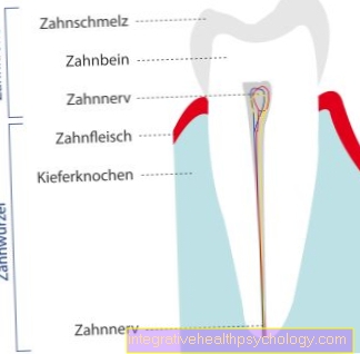
Figure inguinal hernia

- Peritoneal cavity -
Cavitas peritonealis - Abdominal viscera
- Peritoneum -
peritoneum - Glued peritoneum protuberance
- Vas deferens -
Deferens duct - Epididymis -
Epididymis - Testicles -
Testis - Serous testicular envelope -
Tunica vaginalis testis - Scrotum - scrotum
- Inguinal ligament -
Inguinal ligament - Hernial sac
Inguinal hernia - Inguinal hernia
Inguinal hernia types:
a - Epigastric hernia
(in the upper abdomen on the midline) -
Epigastric hernia
b - umbilical hernia -
Umbilical and paraumbilical hernia
c - hernia
(at the location of a previous
surgical intervention) -
Hernia cicatrica
d - Direct inguinal hernia
(in the bar near the
Opening of the inguinal canal)
e - Indirect inguinal hernia
(in the bar at the opening
of the inguinal canal)
f - femoral fracture
(in the thigh canal) -
Femoral hernia
You can find an overview of all Dr-Gumpert images at: medical illustrations
Related images

Illustration
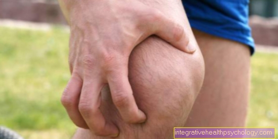
Pain in the
strip

Illustration
Anterior pain
Thigh


.jpg)