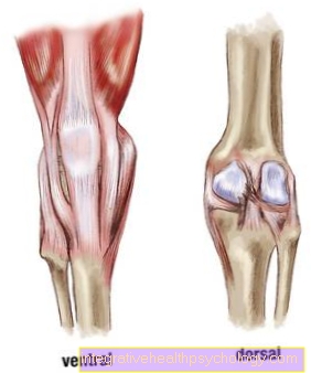
Figure cerebral hemorrhage

Cerebral haemorrhage (cerebral haemorrhage)
- Skull roof -
Calvaria - Hard meninges (dura) -
Cranial dura mater
(outermost meninges) - Subdural gap -
Subdural space - Cobweb skin of the brain -
Arachnoid mater cranialis
(middle meninges) - External cerebral water space -
Subarachnoid space - Cerebrum covered by soft
Meninges (pia) -
Pia mater cranialis
(inner meninges) - bruise
(Hematoma) in brain tissue
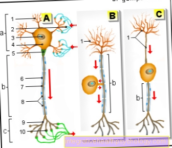
Bleeding in the area of the brain tissue
and the meninges:
A - Intracerebral Haemorrhage (ICB) -
Bleeding into the brain tissue
(Parenchyma) by bursting a cerebral vessel
B - Epidural Bleeding -
Blood between the bones of the skull and the
outermost meninges (Dura mater)
C - Subdural Bleeding -
Blood between the meninges
and cobweb skin
D - subarachnoid hemorrhage -
Blood between cobweb skin and
the inner (soft) meninges
You can find an overview of all Dr-Gumpert images at: medical illustrations
Related images

Illustration
Increased intracranial pressure

Illustration
brain

Illustration
Meninges

Illustration
Brain cysts