Spine anatomy
introduction
The spine represents our "support corset" for walking upright. Ligaments, numerous small joints and auxiliary structures guarantee us not only stability but also a certain amount of flexibility.
Structure of the spine
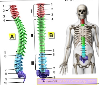
Our spine is divided into the following different sections starting from the head:
- Cervical spine (cervical spine)
- Thoracic spine (BWS)
- Lumbar spine (lumbar spine)
- Sacral spine (SWS)
Figure spine

- First cervical vertebra (carrier) -
Atlas - Second cervical vertebra (turner) -
Axis - Seventh cervical vertebra -
Vertebra prominent - First thoracic vertebra -
Vertebra thoracica I - Twelfth thoracic vertebra -
Vertebra thoracica XII - First lumbar vertebra -
Vertebra lumbalis I - Fifth lumbar vertebra -
Vertebra lumbalis V - Lumbar cruciate ligament kink -
Promontory - Sacrum - Sacrum
- Tailbone - Os coccygis
I - cervical spine (red)
II - thoracic spine (green)
III - lumbar spine (blue)
You can find an overview of all Dr-Gumpert images at: medical illustrations
As a result of the upright, bipedal gait and movement, various curvatures have arisen in these sections as a result of the cushioning and loading, which can be seen from the side. In medicine they are called Lordosis and Kyphosis designated. The former is a protrusion of the spine forward that Kyphosis curves backwards in the side view, like a hump. These special curves are still completely absent in the newborn. They only develop in the course of life. From the birth The predominant continuous backward curvature (kyphosis) is created with the help of the growing neck muscles Neck lordosis to balance the head.
In the further course - with learning the Sitting, Standing and Walking - will the Lumbar lordosis pronounced. These intensify until the legs are in the Hip joints can be stretched, but is only finally fixed in the course of puberty. So there is one in adult humans Cervical lordosis, Thoracic kyphosis, Lumbar lordosis andSacral cyphosis. In the picture there is thus a double S-shaped curvature. From behind, however, you should be able to see a reasonably straight line.
The component of the spine is the individual whirl . In principle, all vortices can be combined into one Vertebral bodies, Vertebral arch and various Appendages (mandrel-, Cross- and Articular process) subdivide. Exceptions here are the 1st and 2nd cervical vertebrae. However, the individual spinal column sections also have special features depending on their function.
In general, the Vertebral bodies and the Vertebral arches the Vertebral hole and in their entirety the Spinal canalwho that Spinal cord houses. The processes that arise from the vertebral arch serve Muscles and Ribbons as an approach. In the area of the thoracic vertebrae, they form the costal vertebrae. There is one between each vertebraIntervertebral disc, the so-called Intervertebral disc.
Appointment with a back specialist?

I would be happy to advise you!
Who am I?
My name is I am a specialist in orthopedics and the founder of .
Various television programs and print media report regularly about my work. On HR television you can see me every 6 weeks live on "Hallo Hessen".
But now enough is indicated ;-)
The spine is difficult to treat. On the one hand it is exposed to high mechanical loads, on the other hand it has great mobility.
The treatment of the spine (e.g. herniated disc, facet syndrome, foramen stenosis, etc.) therefore requires a lot of experience.
I focus on a wide variety of diseases of the spine.
The aim of any treatment is treatment without surgery.
Which therapy achieves the best results in the long term can only be determined after looking at all of the information (Examination, X-ray, ultrasound, MRI, etc.) be assessed.
You can find me in:
- - your orthopedic surgeon
14
Directly to the online appointment arrangement
Unfortunately, it is currently only possible to make an appointment with private health insurers. I hope for your understanding!
Further information about myself can be found at

A - Fifth cervical vertebra (red)
B - sixth thoracic vertebra (green)
C - third lumbar vertebra (blue)
- Vertebral bodies - Corpus vertebrae
- Vortex hole - Vertebral foramen
- Spinous process
(mostly in cervical vertebrae
divided into two) -
Spinous process - Transverse process -
Transverse process - Articular surface for the rib -
Fovea costalis processus - Upper articular process -
Superior articular process - Vertebral arch - Arcus vertebrae
- Articular surface for the rib
on the vertebral body -
Fovea costalis superior - Rib-transverse process joint -
Articulatio costotransversaria - Rib - Costa
- Rib head joint -
Articulatio capitis costae - Transverse process hole
(only for cervical vertebrae) -
Foramen transversarium - Transverse process of the lumbar vertebra
("Costal process") -
Costiform process
You can find an overview of all Dr-Gumpert images at: medical illustrations
Intervertebral discs and ligaments
A Intervertebral disc (= intervertebral disk) represents the cartilaginous connection between two vertebral bodies. It consists of a connective tissue and cartilaginous outer ring, the so-called Annulus fibrosus and a soft inner gelatinous core known as Nucleus pulposus designated.

- Intervertebral disc
(Intervertebral disc) -
Discus inter vertebralis - Gelatinous core - Nucleus pulposus
- Fiber ring - Annulus fibrosus
- Spinal nerve - N. spinalis
- Spinal cord - Medula spinalis
- Spinous process - Spinous process
- Transverse process -
Transverse process - Upper articular process -
Superior articular process - Intervertebral hole -
Intervertebral foramen - Vertebral bodies - Corpus vertebrae
- Front longitudinal ligament -
Lig.longitudinale anterius
The Intervertebral disc takes on the function of a buffer and thus cushions shocks and vibrations that affect the spine. In addition, it enables the individual vertebrae to move better with one another. Not all vertebrae have such a buffer: The first and second cervical vertebrae form a special joint and therefore have a different structure. The same applies to the sacrum and coccyx vertebrae, which merge with one another during development (see: sacrum and coccyx above).
Due to the important tasks and functions that are assigned to the intervertebral disc, it is understandable that it must be shown a special responsibility towards it. This means: Damage to the spine must be avoided if possible. This can be achieved, for example, through "back-friendly" behavior ("Back school“).
In addition, however, it is also of particular importance that the intervertebral disc as such is properly nourished. This "correct" diet has basically nothing to do with healthy food intake as such. The mobility and elasticity of the intervertebral disc is achieved through regular fluid intake, which in turn can only be achieved through healthy and sufficient movement of the person. If the intervertebral disc is alternately loaded and relieved, sufficient fluid absorption is usually guaranteed by “working into the intervertebral disc”.
Nothing is as important as movement for maintaining the elasticity of the intervertebral disc. However, this amount of movement should be appropriate. This means that even permanent movement with only slight breaks can have just as negative effects as a chronic lack of exercise.
In both cases, the cartilaginous outer ring can become brittle and cracked. This gives the inner gelatinous core the opportunity to emerge, so that it may then enter disc prolapse can develop.
So that the spine can be guaranteed maximum support, but also maximum mobility, strong ligaments must be present, which on the one hand extend over the entire length of the spine. In addition, further ligaments are necessary, which will be presented in the course.
- The front longitudinal band is responsible for the stabilization between the abdomen and the spine.
- The rear longitudinal ligament extends over the posterior vertebral body surfaces and lines the anterior vertebral canal area.
- The yellow ribbon (= Ligamentum flavum),
is located between the respective vertebral arches. - A ligament system connects the transverse processes of the individual vertebrae with the intermediate transverse processes.
- A ligament system (= intermediate spinous process ligaments) connects the spinous processes and thus the back of the vertebrae.
- A ligament also extends over all spinous processes and supports the spine in the form of posterior stabilization.
The Back muscles also provides additional support for the entire belt system. Only the joint effect and mutual support enable the well-known elastic and stabilizing function and structure of the spine and thus enable the numerous possibilities of movement in all directions, including any rotational movements.
Band washers

The intervertebral disc acts as a buffer between two vertebrae. It consists of an outer fiber ring (Annulus fibrosus) and an inner core of gel-like mass (Nucleus pulposus). The core is used for reversible water binding, i.e. it can - depending on the current load condition of the respective spinal column segment - release water (heavy load) or absorb water (decreasing load), it thus acts like a kind of water cushion or sponge.
The intervertebral disc is the shock absorber of the spine and is therefore exposed to enormous forces, which is reflected in the increasingly common disc bulges or even prolapsed disc protrusions in today's patient population. In such a herniated disc, the outer fiber ring becomes porous and cracked, so that parts of the core emerge and partially slide into the spinal canal, where they can irritate nerves running there (see below).
Tape apparatus
Numerous ligaments stabilize the bony spine. These include the anterior and posterior longitudinal ligament (Lig.longitudinale anterius and posterius) running along the entire spine of cranial to caudal run, the yellow ribbons (Ligamenta flava), which connect the adjacent vertebral arches and the ligaments between the spinous processes (Ligamenta interspinalia).
More information can be found here: The ligaments of the spine
Spinal cord
The spinal cord runs through the spinal canal, which is connected by the individual vertebral holes (Vertebral foramina) is formed caudally and gives off a nerve cord (the spinal nerve) to the right and left on each vertebral body. This spinal nerve runs through the intervertebral holes (Intervertebral foramina) and thus leaves the spinal canal.
There are 31 pairs of spinal nerves. 8 cervical (belonging to the cervical spine), 12 thoracic (belonging to the thoracic spine), 5 lumbar (belonging to the lumbar spine), 5 sacral (belonging to the sacrum / sacral spine) and 1 coccygales (belonging to the tailbone), which in humans is only rudimentary is.
The first spinal nerve occurs in the area of the cervical spine (C1) above the first cervical vertebra (HWK 1), so that the spinal nerve emerges in the area of the cervical spine above its associated vertebral body. However, the fact that there are 8 cervical spinal nerves and only 7 cervical vertebrae changes this pattern with the 8th spinal nerve, which emerges below the 7th cervical vertebrae.
The 1st thoracic spinal nerve (Th 1) below its associated vertebral body (BWK 1).
The spinal cord as such ends at the level of the 1st lumbar vertebral body, while the spinal nerves run even further down on the way to the outlet openings assigned to them. This bundle of spinal nerves, which however no longer includes the spinal cord itself, is called Cauda equina (in German: ponytail). When taking cerebral fluid in the area of the back (lumbar puncture or CSF puncture), a needle can be inserted from the 2nd lumbar vertebra (but usually between the 3rd and 4th lumbar vertebrae) without the risk of injuring the spinal cord. The cauda equina running there is flexible and can evade the tip of the needle.
annoy
The spine forms a bony protective wall around the human spinal cord through which the nerve cords run electrical impulses to the muscles send. Also sensitive sensory perceptions are from the periphery via the Spinal cord directed to the brain, where they can be consciously perceived. In order to get to the peripheral areas of the body, for example the arms and legs, nerve cords pull out of the spinal cord between the individual vertebral bodies.
For any damage to the spine, for example Vertebral fractures, Herniated discs and degenerative spinal diseases, the nerves in the spine are at risk due to their proximity. In the case of pain that occurs in the back and spreads to the periphery, there may be nerve involvement that needs urgent treatment.
The spinal cord itself, which runs within the spinal column in the spinal canal, consists of Nerve tissue. In cross-section, the spinal cord appears as an approximately round, bright surface (white matter), in the middle of which there is a butterfly-shaped darker, gray structure (gray matter). While the gray matter is formed by the bodies of the nerve cells (perikaryen), the white area around them represents their projections (axons).
The The spinal cord contains different pathways with different qualities that convey information from the brain to the rest of the body (the periphery) and from the periphery back to the brain. Movement commands are passed from the brain to the muscles or, conversely, perceptions such as pain are passed from the skin to the brain. The spinal cord is as essential as that Mediator between the brain and the rest of the body.
Two vertebral bodies lying one below the other form an intervertebral hole (foramen intervertebrale) through which the spinal nerves emerge. 31 pairs of these arise directly from the spinal cord, but belong to the peripheral nervous system. They are all mixed nerves, thus contain sensitive (e.g. feeling or pain perception), motor (movement) and vegetative (e.g. sweating) qualities.
Nerve root
Nerve roots are fibers that go in or out of the spinal cord. On each section of the spine (segment) there are 2 on the right and left Nerve roots, one Rear and a Front.
The front roots conduct motor commands from the brain to the muscles, whereas the Back sensitive information like pain or touch direct from the body to the brain. The 2 roots of one side unite in the spinal canal to form a spinal nerve (spinal nerve). A spinal nerve leaves the spinal canal through an intervertebral hole on each side.
Cervical spine

Of the total of 7 cervical vertebrae, the first (atlas) and second (axis) deviate the most from the basic shape of the vertebrae. They are built in such a way that they can both bear the main load of the head and enable movement in three degrees of freedom, similar to a ball joint. The first cervical vertebra “atlas”, named after Greek mythology, lies directly under the occipital opening (foramen magnum) of the skull, carries its entire load and includes the tooth of the second cervical vertebra, the axis. The other five cervical vertebrae (cervical spine) have a relatively small vertebral body, which is almost cube-shaped when viewed from above, and a large, triangular vertebral hole in which the nerve pathways coming from the skull continue as the spinal cord. As an anatomical peculiarity, the transverse processes of the cervical spine are split and thus form a channel that leads to an artery supplying the brain (arteria vertebralis) on the left and right. The upper surface of the transverse process has a deep, wide groove from the 3rd cervical vertebra through which the respective spinal nerve emerges outward through the intervertebral hole. Eight bundles of nerves arise on each side in the area of the cervical spine. The top four form the cervical nerve plexus, which innervates the neck muscles and the diaphragm, the most important respiratory muscle.
In the event of an injury above these spinal cord segments, e.g. as a result of a car accident, independent breathing is no longer possible. The lower four nerve bundles together with the first of the thoracic spine form the arm nerve plexus, which is responsible for the motor functions of the arm and chest muscles as well as the skin areas in these areas.
The seventh cervical vertebra can be quickly identified from the outside through the backward protruding spinous process. This gave it its own name: Vertebra prominens.
The articular processes articulate the individual vertebrae with one another upwards and downwards.
Read more on the topic: Cervical spine
Thoracic spine
The Thoracic spine consists of 12 vertebrae. The vertebral bodies gradually become higher and wider as they progress towards the lumbar spine. The vertebral hole is approximately round and smaller than in the cervical and lumbar spine, the end surfaces are rounded and triangular. Since the spinous processes are long and sharply bent back and down, the thoracic vertebrae are connected in a special way (like Roof tiles) toothed. To the Thoracic vertebrae put the Ribs that is why they are equipped with articular surfaces covered with cartilage both on the vertebral bodies and on the transverse processes. So there are two Rib-vertebral joints: the Rib head joint and the Costal hump joint.
The former is used in the 2nd-10th rib formed by two vertebral bodies standing on top of each other and the head of the rib with their joint surfaces.
In the 1st, 11th and 12th rib only articulates one Thoracic vertebrae with the rib head. All joint capsules of the rib head joints are reinforced by ligaments.At the rib cusp joints of the 1-10 rib articulate the rib cusps with the articular surface of the corresponding thoracic vertebral process.
In the 11th and 12th rib there is no corresponding joint because the transverse processes of these thoracic vertebrae have no articular surfaces. These joints are also reinforced by a total of 3 ligaments. They run not only between the ribs and their associated thoracic vertebrae, but also between the rib neck and the transverse process of the next higher vertebra.
Both rib joints are morphologically completely separated from one another, but their mobility forms a unit.
Lumbar spine

In the lumbar spine, the rib structures in the form of the transverse processes are much more powerful than in the cervical spine. Therefore, the transverse processes in this area are also referred to as rib processes. Additional ribs can occur, but they usually do not cause any discomfort. On the other hand, if there is an additional cervical rib, the arm nerve plexus and the accompanying artery can be constricted and the so-called scalenus or cervical rib syndrome occurs.
The lumbar spine has 5 strong vertebral bodies that are transversely oval when viewed from above. Their massive vertebral arches enclose an almost triangular vertebral hole and unite to form a strong, flattened spinous process. Due to the upright gait, there is an enormous weight on the lumbar spine. This stress can lead to various clinical pictures. From unspecific pain to degenerative changes to the dangerous herniated disc, which is often found in this area, the lumbar spine is particularly in the focus of clinicians.
Inside the spinal canal there is a special feature of the lumbar spine and the spinal cord that runs in it.
In most people, this ends at the level of the 2nd lumbar vertebra. This fact goes back to the history of human development. Up to the 12th week of development in the womb, the spinal cord and spinal canal are of the same length, so that the pair of spinal nerves emerge through the intervertebral hole at the same height. With age, however, the spine grows faster than the spinal cord, so that the spinal cord ends at the level of the 3rd lumbar vertebra at birth. The consequence of this different growth is that the spinal roots of the nerves pull obliquely downward in the spinal canal down to their respective intervertebral hole and exit there. As a whole, these roots are called the so-called "ponytail" (Cauda equina). Although there are no more spinal cord segments in this area, the sheaths or skins surrounding the spinal cord continue to extend into the sacral canal. This is the reason why cerebrospinal fluid in this area is safe (Cerebral and spinal fluid) can be taken. This lumbar puncture is used to diagnose various diseases. An anesthetic can also be used in this area as part of a surgical procedure to eliminate pain and paralyze the muscles in the lower extremities and the pelvic region (spinal lumbar anesthesia).
Read more on the topic: Lumbar spine
Sacral spine
The so-called Sacrum originally consists of five independent vertebrae. After birth, however, these merge uniformly to form a triangular-looking bone when viewed from the front. Nevertheless, the sacrum still has all the characteristics of a vertebra. The fused vertebrae form four T-shaped bone canals in the upper area through which the sacral nerves emerge. The combined spinous processes form a serrated bone ridge on the convex posterior side. On either side of it, the merging of the transverse processes with the rib rudiments on both sides of the sacrum creates the powerful lateral parts that carry ear-shaped joint surfaces for the iliac bones of the pelvis on their sides.
The tailbone joins the sacrum with three to four rudiments of vertebrae. At least the first coccyx vertebra usually still shows typical components.
Ligamentous apparatus of the spine
The Spinal ligaments lead to a stable connection between the vertebrae and enable high mechanical loads. Vertebral body ligaments and vertebral arch ligaments can be distinguished from one another within the ligamentous apparatus.
The anterior vertebral ligament runs across the front of the vertebral bodies from the base of the skull to the sacrum. With its deep fibers it connects neighboring vertebral bodies, with its superficial parts it extends over several segments. This ligament is only loosely connected to the intervertebral discs. The posterior vertebral ligament runs from the posterior fossa over the back of the vertebral bodies and into the sacral canal. In contrast to the anterior ligament, the posterior ligament is firmly attached to the intervertebral disc. Both ligaments are involved in maintaining the curvature of the spine.
As the name suggests, the vertebral arch ligaments run between the vertebral arches, between the spinous and transverse processes and thus create additional stability.
Range of motion of the spine
For the mobility of the spine they are Vertebral arch joints (so-called small vertebral joints) responsible. They are formed by the articular processes of the vertebral arches and are created in pairs. Since they are tilted against the horizontal to different extents depending on the section of the spine, they have a certain amount of movement and special directions of movement (see table). The following movements are generally possible:
- Forward flexion (Ventral flexion)
- Backward flexion (Dorsiflexion)
- Sideways flexion (Lateral flexion)
- rotation (Rotation)
The following table shows the degree of mobility in the individual sections of the spine:
Cervical spine (Cervical spine):
- Forward flexion: 65 °
- Backward flexion: 40 °
- Sideways flexion: 35 °
- Rotation: 50 °
Thoracic spine (BWS):
- Forward flexion: 35 °
- Backward flexion: 25 °
- Sideways flexion: 20 °
- Rotation: 35 °
Lumbar spine (lumbar spine):
- Forward flexion: 50 °
- Backward flexion: 35 °
- Sideways flexion: 20 °
- Rotation: 5 °
Cervical spine + thoracic spine + lumbar spine:
- Forward flexion: 150 °
- Backward flexion: 100 °
- Sideways flexion: 75 °
- Rotation: 90 °
Function of the spine
The spine is an elaborate structure of the human body that many functions enables.
First of all, she holds that Body upright and is therefore not called the "backbone" for nothing. A complex one Interplay of bony structures, Ligaments and muscles allows the trunk, neck and head to be stabilized. In this respect, humans differ from other vertebrates in their upright gait.
The spine carries the skull upwards and at the same time allows the head to move freely on all sides. Furthermore, the spine is connected to the ribs via many small joints and is related to the shoulder girdle. The sacrum as the lower end of the spine contributes to the formation of the pelvis by forming the so-called pelvic ring with other bones.
Another important function of the spine lies in the fact that they are a bony protection around the delicate spinal cord offers. The spinal cord enters through a bony opening in the skull bone and then runs through the Spinal canal or spinal canal (Canalis vertebralis), which is formed by the individual vertebral bodies lying on top of one another. From the spinal canal there is an opening on both sides, the intervertebral hole (foramen intervertebrale). This is always formed by two vertebrae lying one below the other and is the exit point for the so-called spinal nerves (spinal cord nerves)









-de-quervain.jpg)

