bone
Synonyms
Bone structure, bone structure, skeleton
Medical: Os
English: bone
introduction
The bony skeleton of an adult consists of around 200 individual bones that are connected to one another by joints. They make up about 10% of the body weight. The shape of the individual bones is genetically determined, the structure depends on the type and size of the mechanical load.
The bone substance is arranged in such a way that the greatest possible strength is achieved with the least amount of material. Bones are distinguished on the one hand by shape and on the other hand independently of shape.
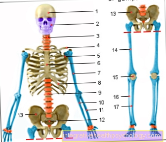
Figure bone overview

Long bones
(Long bones) - blue
Short bones - orange
Plate bone - yellow
Mixed forms - purple
- Brain skull -
Neurocranium - Facial skull -
Viscerocranium - Cervical vertebrae -
Vertebrae cervicales - Collarbone -
Clavicle - Shoulder blade - Scapula
- Sternum - sternum
- Humerus - Humerus
- Ribs - Costae
- Lumbar vertebrae -
Vertebrae lumbales - Cubit - Ulna
- Spoke - radius
- Sacrum - Sacrum
- Hip bone - Os coxae
- Femur - Femur
- Kneecap - patella
- Fibula - Fibula
- Shin - Tibia
You can find an overview of all Dr-Gumpert images at: medical illustrations
Bone shapes
A distinction is made according to the shape:
- long bones
- short bones
- flat planar bones
- irregular bones
Regardless of the shape, a distinction is also made:
- aerated bones
- Sesame legs and additional, so-called
- accessory bones
Appointment with Dr.Gumpert?

I would be happy to advise you!
Who am I?
My name is I am a specialist in orthopedics and the founder of and work as an orthopedist at .
Various television programs and print media report regularly about my work. On HR television you can see me live every 6 weeks on "Hallo Hessen".
But now enough is indicated ;-)
In order to be able to treat successfully in orthopedics, a thorough examination, diagnosis and a medical history are required.
In our very economic world in particular, there is not enough time to thoroughly grasp the complex diseases of orthopedics and thus initiate targeted treatment.
I don't want to join the ranks of "quick knife pullers".
The aim of all treatment is treatment without surgery.
Which therapy achieves the best results in the long term can only be determined after looking at all of the information (Examination, X-ray, ultrasound, MRI, etc.) be assessed.
You will find me:
- - orthopedic surgeons
14
You can make an appointment here.
Unfortunately, it is currently only possible to make an appointment with private health insurers. I hope for your understanding!
For more information about myself, see - Orthopedists.
The long bones of the extremities are Long bones and will be out a shaft (Diaphysis) and two ends (Epiphyses) educated. There is one during the growth phase Growth plate (Epiphyseal plate) out cartilage between the shaft and the epiphysis, which ossifies at the end of the growth phase to form what is known as the epiphyseal plate.
The part of the shaft directly adjoining the epiphyseal plate is called Metaphysis designated. Bone protrusions to which tendons and ligaments are attached are called Apophyses designated. When the tendons and ligaments attach to roughness, this roughness is called Tuberosity designated.
Comb-shaped or ridge-shaped bone edges are called Comb (Crista) or lip (Labrum) or linear roughness (Linea )designated. These combs, lips and lines are used Muscles, Tendons, ligaments and joint capsules as an attachment.
Illustration showing bone structure

a - epiphysis
(Bone end)
b - metaphysis
(active growth zone)
c - diaphysis
(Bone shaft)
- Sponge-like built
Bone with red
Bone marrow -
Substantia cancellous
+ Medulla ossium rubra - Epiphyseal line -
Epiphysial line - Dense (compact) bone -
Substantia compacta - Medullary cavity with yellow
Bone marrow -
Cavitas medullaris
+ Medulla ossium flava - Bony artery -
Nutrician artery - Periosteum -
Periosteum - Osteon (basic functional unit) -
Osteonum - Spaces filled with bone marrow
between the trabeculae -
Medulla ossium - Growth plate -
Lamina epiphysialis
You can find an overview of all Dr-Gumpert images at: medical illustrations
Composition of the bone

The bone tissue consists of the Bone cells (Osteocytes) by a Extracellular matrix out:
- Basic substance
- collagen fibrils
- one Putty substance and
- different Salt is formed.
The Basic substance and the collagen fibrils are also called intercellular substance designated. The collagen fibrils belong to the organic Part of the bone and the salts belong to the inorganic Part. The most important salts in bones are:
- Calcium phosphate
- Magnesium phosphate and
- Calcium carbonate,
the less important are other compounds of calcium, potassium, sodium with chlorine and fluorine. The salts determine the hardness and strength of the bone. If the bone is free of salt, it becomes pliable.
The organic components of the bone ensure elasticity. The ratio of salts and organic components changes in the course of life. In the newborn, the proportion of organic parts of the bone is 50%, only with the old man 30%. In addition to the Osteocytes still to come Osteoblasts as a bone building and Osteoclasts as bone-degrading cells.
The bone tissue is after Dental tissue the hardest substance in the human body and has a water content of 20%.
Formation of the bone
Become bones in two different ways formed in the human body.
- In the desmal bone development (ossification) the bone is formed directly, while in the
- chondral bone development of the bones arises indirectly from cartilage tissue.
In both cases the first bone appendages appear in the 2nd embryonic month with the Collarbone and ends at the end of the Apo- and Epiphyseal plates at the beginning of the 20th year of life. If the bone is located directly in the embryonic connective tissue (Mesenchyme) developed from mesenchymal precursor cells is called desmal bone development.
The resulting bones are called Connective tissue bones designated. This is how, among other things Skull bones, the Lower jaw and parts of the Collarbone.
If the bone does not develop from the connective tissue, but from cartilage tissue, one speaks of a chondral ossification. First of all, a cartilaginous pre-skeleton (Primary skeleton), which is similar in shape to the later skeleton. This “pre-skeleton” is then replaced by bones. Both forms initially lead to the formation of braided bone, which then transforms into the lamellar bone under load. The braided bone has a greater growth potential than the lamellar bone and thus forms more Afford and Trabeculae with the help of which he can erect extensive scaffolding in a relatively short time.
Within the woven bone, the blood vessels and the course of the collagen fibers are disordered and the number of osteocytes is small and their arrangement is random. In addition, the mineralization content of the tissue is low. This is why the braided bone is not as resilient as the lamellar bone. During growth well into the 1920s, the braided bone is transformed into lamellar bone. The first generation of the resulting osteons are called primary osteons and arise during the Fetal time.
When these are replaced by new osteons, which are now referred to as secondary osteons, through remodeling processes. This remodeling takes place increasingly between the 8 and 15 years of age instead of. During the remodeling, vessels first penetrate the woven bone and, with the help of osteoclasts, drive a vascular canal into the bone. This already has the diameter of the future osteon. The osteoblasts then differentiate from the connective tissue accompanying the vessels, attach to the canal wall and begin to form the matrix, which is already arranged as an osteoid in the form of lamellae in the osteon.
Later, the osteoid is completely mineralized and the osteoblasts are walled in. The lumen of the canal is narrowed bit by bit until only the Haversian canal remains.
Development of the long bone
Developing a Long bone runs through both the direct read and the indirect ossification. The so-called perichondral bone cuff is created within the bone shaft via direct ossification. On its basis, the shaft grows in thickness. Additional fiber and braided bone trabeculae are attached to the perichondral bone cuff until a loosely structured bony shaft has formed. For the time being, the ring only forms in the central shaft area, but then extends over the entire length of the shaft. This leads to a stiffening and the further bony remodeling processes do not lead to an interruption of the support function.
With the appearance of braided bones, the temporary surrounding one becomes Perichondrium converted to the periosteum, from which the further growth of the bone proceeds. This is followed by strong cartilage growth in the area of the shaft, which provokes a growth in length of the shaft. Here the cartilage cells are already arranged in longitudinal cell columns, which then ossify.
Due to a deteriorated supply of the cartilage cells with nutrients, these are then broken down by connective tissue penetrating from the vessels with the help of cartilage-degrading cells. This creates a primary medullary cavity in which the Bone marrow with its mesenchymal cells.
At the edges of the Medullary cavity Then osteoblasts begin to form bone mass, so that a primary bone core is created. Starting from the primary medullary cavity, the cartilage is then gradually replaced by the woven bone down to the epiphyses. At a genetically determined point in time, secondary bone nuclei arise within the epiphysis, which then displace the cartilage tissue from the epiphyses. At the epiphyseal plates the cartilage is increased by division and this is how the length increases. Through a cartilage plate is the bony epiphysis of the Metaphysis Cut. The articular cartilage is connected to the growth zone. Four zones are distinguished within the epiphyseal plate,
- the Reserve zone (with resting cartilage),
- the Proliferation zone (with columnar cartilage cells),
- the Cartilage remodeling zone and the
- ossification.
The proliferation zone is crucial for long growth. This is where the cells multiply. Characteristic cell columns are created through cell division. As the cells increase in size, they take up more water and are then located in the bladder cartilage zone. This cell hypertrophy and cell division benefit the growth in length. Cell activity increases in the bladder cartilage zone, increased collagen formation occurs, which form longitudinal septa and mineralization, so that stiffening occurs. This is a prerequisite for the growth of vessels and the septa serve as a framework for the newly formed bone.
Cartilage-eating cells enter the tissue via the vessels and build the cartilage, so that space is created for the bone that is formed. Bone formation then begins with osteoblast colonization on the surface of the remaining mineralized septa.









.jpg)

