Ledderhose's disease
synonym
Plantar fascial fibromatosis
definition
Ledderhose's disease is one benign disease of the connective tissue of the feet. It occurs in the area of the plantar aponeuroses (= Latin term for the tendon plate of the sole of the foot). More precisely, it is a thickening of the deep connective tissue or fascia in the foot. Ledderhose's disease is one of the symptoms of Fibromatoses, and is also associated with the Dupuytren's disease related, which is a benign connective tissue disease of the palm of the hand.
The nodes on the soles of the feet usually grow very slowly and are almost always centered on the plantar fascia (sole of the foot). Occasionally the nodes will slow down and stop enlarging. Then suddenly they can grow rapidly and unexpectedly again. Surgical intervention is only necessary for painful lumps that impede walking.

causes
The causes of the disease are not yet fully understood. It is known that the protrusion on the sole of the foot is caused by a Increase in connective tissue arise in the affected area. More precisely, certain cells, the myofibroblasts, are responsible for this.
There are a number of different theories and conjectures which factors can influence the occurrence of Ledderhose's disease. So it is considered likely that a genetic component plays a role in the disease. The connective tissue changes occur when external influencing factors such as injuries or other events of an unknown nature are added. Another argument in favor of a genetic influence is that Men about twice as often are more affected than women.
Other risk factors are the simultaneous presence ontheir fibromatoses - especially at Dupuytren's disease - as well as certain diseases like Diabetes mellitus or one Epilepsy.
There are also a number of different factors, the significance of which in the development of the disease has not yet been proven, but indications of this have appeared in individual cases. This includes in particular the consumption of luxury foods such as nicotine and alcohol such as stress, and certain Metabolic and liver diseases.
Appointment with ?

I would be happy to advise you!
Who am I?
My name is I am a specialist in orthopedics and the founder of .
Various television programs and print media report regularly about my work. On HR television you can see me every 6 weeks live on "Hallo Hessen".
But now enough is indicated ;-)
Athletes (joggers, soccer players, etc.) are particularly often affected by diseases of the foot. In some cases, the cause of the foot discomfort cannot be identified at first.
Therefore, the treatment of the foot (e.g. Achilles tendonitis, heel spurs, etc.) requires a lot of experience.
I focus on a wide variety of foot diseases.
The aim of every treatment is treatment without surgery with a complete recovery of performance.
Which therapy achieves the best results in the long term can only be determined after looking at all of the information (Examination, X-ray, ultrasound, MRI, etc.) be assessed.
You can find me in:
- - your orthopedic surgeon
14
Directly to the online appointment arrangement
Unfortunately, it is currently only possible to make an appointment with private health insurers. I hope for your understanding!
Further information about myself can be found at
Symptoms
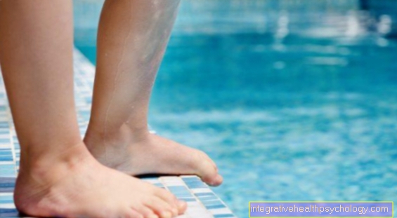
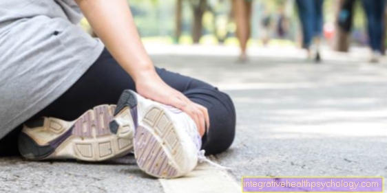
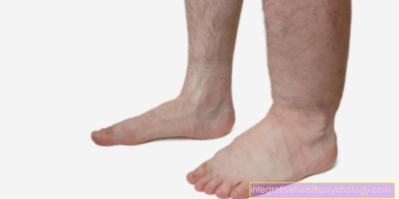
In Ledderhose's disease is usually the Impaired walking ability. This is because the nodes are on the sole of the foot, especially at the highest point of the arch in the middle of the sole. There can be just one knot or multiple knots as well Strand formation. If these are pronounced and distributed over the entire sole of the foot, the knots are mostly fixed to the Musculature and the one above skin grown together. In contrast, in the mild form of Ledderhose's disease, only a small part of the plantar fascia is affected and neither skin nor muscles show adhesions. Around 25% of those affected have Ledderhose's disease on both feet.
diagnosis
At the beginning of the diagnosis of Ledderhose's disease there is the anamnese of the person concerned. The attending physician can often make the suspected diagnosis of Ledderhose's disease due to the symptoms typically occurring when walking, which the person concerned often first notices, as well as the physical examination.
When examining the foot fall relative hard knots that are difficult to move by hand. Come to be able to determine the actual size of the knot imaging diagnostic Devices used. Especially performing one Ultrasound examination can be carried out in many general practices. To get a more precise picture of the individual spread of the nodes, pictures with a Magnetic resonance tomograph (MRI) to be created.
Absolute certainty about the presence of Ledderhose's disease allows microscopic Examination of the nodes. The examined material can on the one hand by a biopsy or removed during surgery to remove the nodes and examined by a pathologist.
Imaging
in the Magnetic resonance imaging The typical Ledderhose's disease shows up as a poorly defined, infiltrative mass in the tendon plate near the plantar muscle.
MRI of the foot
To rule out possible differential diagnoses of nodular changes in the foot is one magnetic resonance imaging examination, i.e. an MRI of the foot. The MRI is particularly suitable for visualizing soft tissue. Since the nodular changes in the Ledderhose muscle are connective tissue cell material, this is a space occupation based on the tendon plate of the foot (Plantar fascia) can be seen in the MRI. The Signal intensity can be assessed in different sequences. In the possible sequences, the connective tissue-like change is low in signal compared to the surrounding tissue, i.e. dark. It can also be seen that the fibromatous structure infiltrating grows, i.e. pulls into surrounding structures such as muscles, tendons, fat and skin. If contrast agent is also injected, there is also a even contrast enhancement of watching.
Further information can be found on our website MRI of the foot.
Which doctor should I go to?
Usually will when complaints arise for the first time or the symptom-free knowledge of the tumor formation on the sole of the foot Family doctor visited because the layman usually does not know what this connective tissue change can be. Depending on experience and the availability of imaging equipment (ultrasound), the family doctor can make the diagnosis himself.
To more detailed clarification can he also do a Referral for an MRI to a radiologist (X-ray doctor), who can ultimately secure the diagnosis with the help of the images. The family doctor can also be consulted for therapeutic, conservative measures.
Depending on further treatment, the nodular change surgically removed by a surgeon become. This is mostly about Foot surgeonswho carry out the procedure on an in-patient basis, but often also on an out-patient basis. Since foot surgery is a specialty, it is advisable to have the operation performed in a specialist clinic.
therapy
An important guideline in the treatment of Ledderhose's disease is to reduce inflammation and pain, and to maintain the patient's ability to walk.
Soft pads can be prescribed that can prevent internal pressure on the nodes. For the inflammation and pain, non-steroidal anti-inflammatory drugs are often prescribed, as well as steroid injections into the nodes.
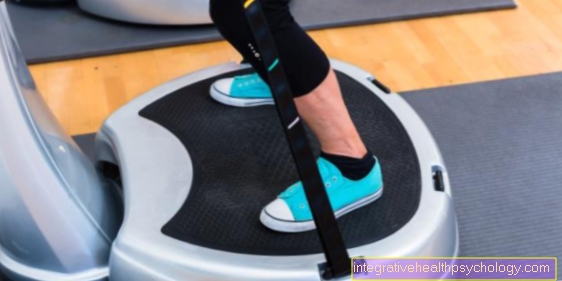
Radiotherapy with soft X-rays often shows good results in the early stages. Furthermore, therapy with shock waves or the injection of collagenases, which are supposed to loosen the hardened knots, have also brought good results.
In the case of existing symptoms and in an advanced stage, Ledderhose's disease must be operated on. Radical removal of the plantar fascia is often recommended, as even faster-growing lumps often reappear with minimal intervention. However, it must also be explained to the person concerned that the possibility of a return of the fibromatosis is 25%. In addition, the risks of an intervention on the sole of the foot must be explained. Nerves, muscles and vision are close together here and could be injured.
Irradiation
The use of radiation therapy in the treatment of Ledderhose's disease is mainly found in Early stages great attention. Some studies have shown the effectiveness of radiation therapy.
A distinction must be made between two different forms of radiation for the radiation used. It'll come once mild x-rays (Orthovolt therapy) as well Electron beams for use.
The radiation energy in these treatments for Ledderhose's disease is only a fraction of that which is used, for example, for malignant, solid tumors. Nevertheless, there is a certain risk for the person being treated, which is why usually only People over 45 years of age receiving radiation therapy.
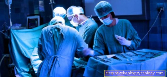
surgery

The therapy options for Ledderhose's disease can be found in conservative such as operational Treatment options are classified. If conservative methods do not show any therapeutic success, an operation can be considered.
There are two different ways of operating the nodes on the sole of the foot. For one thing, only the Knot removed become. This initially frees you from symptoms, but is associated with a high probability that further, even more aggressive and larger lumps will develop over time. The probability that with this type of removal Recurrences arise is attached up to 85%.
The second option is to remove the nodes and do so at the same time Removal of the so-called plantar fascia (Plantar fasciactomy). This fascia is a tendon plate which is located on the sole of the foot and is the starting point for the formation of the nodes. But even after the plantar fascia has been removed, recurrences can occur. The likelihood that after performing this surgery Recurrences arise is attached about 25%.
Since the likelihood of recurrence is significantly lower, many doctors recommend the latter when choosing between surgery. This is also justified with the fact that the recurrences that occur are a more aggressive growth and a second operation has a higher risk of complications due to the scar tissue that has formed.
However, it should be mentioned that removing the plantar fascia is not without consequences for the affected person. So can others Discomfort when walking occur which the attending physician must inform about before the operation is carried out.
If the skin is so severely damaged by the connective tissue that it has to be removed over a large area, a Skin graft to the sole of the foot.
In both operations, the affected foot should be used for spared for up to three weeks become. This is necessary on the one hand to allow the wound to heal as quickly as possible and to reduce the likelihood of recurrences somewhat.
homeopathy
In addition to classic treatment approaches such as conservative and / or surgical care and radiotherapy, homeopathy is becoming more and more popular. This continues with different homeopathic remedies especially the Pain and inflammation relief as goal.
One substance that is supposed to help in the homeopathic treatment of Ledderhose's disease is that Formic acid (Acidum formicicum). The application provides for the formic acid in the area of the plantar fascia, ie at the place of manifestation, to be injected. This procedure is described by those treated as very painful, but it must take place several times for a successful therapy. However, there are currently no studies that confirm the benefits or effectiveness of homeopathic treatment. Therefore, if the therapeutic benefit is inadequate and the pain is too severe during the use of formic acid injections, those affected often resort to medicinal or surgical treatment.
cure
M. Ledderhose is a benign connective tissue overgrowth, which treated with different therapeutic approaches can be. Conservative treatments make it possible Prevent progression of the nodular growths or even to eliminate it entirely.
However, M. Ledderhose has the property to occur in bursts and a progressive (= advancing) course to pursue. This means that even after successful therapy and after phases of symptom relief, a new flare occurs and the nodular changes become symptomatic again. Also the surgical removal can no lifetime guarantee to ensure that the disease does not break out again. As with the analogous clinical picture of Dupuytren's disease, the recurrence rate is very high.
Risk factors
Why even one Ledderhose's disease occurs, is unfortunately not yet exactly known. There are currently defined risk factors that favor the occurrence of plantar fascial fibromatosis.
Which includes:
- familial frequency of the disease
- Gender (men are more often affected than women)
- Fibromatosis in hand (this increases the risk to 10-65%)
- Induratio penis plastica disease
- epilepsy
- Diabetes mellitus
Other factors whose clear connection has not yet been proven: smoking, alcoholism, Liver disease, Thyroid disease, Stress.
Analogies to Dupuytren's disease
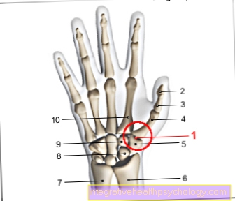
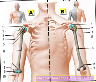
The disease picture M. Ledderhose counts just like that Dupuytren's contracture to Group of benign connective tissue growths, called fibromatosis. Ledderhose's disease is a connective tissue disease of the tendon plate (Aponeurosis) of the feet, the plantar fascia. Similarly, the disease on the hands is called Dupuytren's disease and affects the hands there Tendon plate of the handwho have favourited Palmar Aponeurosis.
Both is togetherthat it is a benign, connective tissue growth which can grow into the surrounding tissue and is largely based on the multiplication of special cells, the so-called myofibroblasts. In addition, both have diseases a high risk of recurrence after surgical removali.e. that even after complete removal, the nodular change can always reappear.
A third related disease concerns the limb and is called "Induratio penis plastica“, A scarring of certain layers of the skin, which is associated with a painful curvature of the penis during erection and the risk of erectile dysfunction.
Of the 3 named fibromatoses, the following applies Dupuytren's contracture as the most common and most well-known clinical picture. Despite the many similarities, M. Ledderhose and M. Dupuytren differ in a few ways. On the one hand, there is one in Dupuytren's disease Extension inhibition of the fingers, hence the synonym Dupuytren's contracture (contracture = shortening of muscles and tendons). However, this symptom does not occur on the foot, since the toes are usually not affected to this extent there. On the other hand, the nodular changes on the plantar fascia of the foot tend to be a lot larger than those on the palmar fascia of the hand.
Georg Ledderhose
The German doctor Georg Ledderhose (1855-1925) discovered and described the disease. Furthermore, the surgeon who works in Strasbourg and Munich had that Glucosamine discovered. Glucosamine is an important component of the synovial fluid and cartilage.