artery
Synonyms
Artery, artery, pulse artery, artery, blood vessel, vessel
English: artery
definition

An artery is a blood vessel that carries blood away from the heart. In the body's circulation, an artery always carries oxygen-rich blood, while in the pulmonary circulation it always carries oxygen-poor blood, as it transports the oxygen-poor blood from the heart to the lungs for oxygenation. Depending on their diameter and position in the body, arteries change their microscopic (histological) Construction. A distinction is also made between small arteries, so-called arterioles, and the smallest hair vessels, the so-called capillaries.Compared to veins, arteries are thicker-walled, as there is a high internal pressure (blood pressure) in arteries and this is counteracted by this. Furthermore, arteries have a round inner shape (Lumens).
The arteries are the high pressure system of the blood system. The arterial internal pressure varies between the ejection phase (systole), i.e. the maximum contraction of the heart, and the filling phase (diastole) of the heart.
The largest artery in the human body is the main artery (aorta). It has a diameter of up to three centimeters, depending on the body shape.
Illustration of an artery

- Outer layer of
Arterial wall -
Tunica externa - Outer elastic layer -
External elastic membrane - Middle layer of the arterial wall -
Tunica media - Inner elastic layer -
Membrana elastica interna - Inner layer of the arterial wall -
Tunica intima - Endothelial cells - Endotheliocyti
- Blood vessels in the adventitia -
Vasa vasorum - Autonomous nerve network of the
Vessel wall -
Vascular plexus
You can find an overview of all Dr-Gumpert images at: medical illustrations
Microscopic wall structure
An artery is made up of three layers. The first and innermost layer, i.e. the layer that comes into contact with the blood flowing through, consists of a single layer of cells, a so-called single-layer uncornified squamous epithelium. This innermost layer is also known as the endothelium or intima (Tunica intima). It is the crucial barrier between the inside of the blood vessel (intravascular space), i.e. the blood, and the area outside the blood vessel (extravascular space).
The second following layer consists mainly of smooth, non-arbitrarily controllable muscles that cannot be controlled arbitrarily. This layer is called media (Tunica Media). In addition to the smooth muscles, the second layer also contains elastic fibers, depending on the type in the body. This layer is mainly used to adjust the wall tension of an artery and the vessel width. When the smooth muscles contract, the wall tension increases and the artery becomes narrower.
The third and outermost layer of an artery is called the adventitia (Tunica adventitia). The adventitia consists mainly of connective tissue, which anchors the artery with the environment in the body. Furthermore, the adventitia determines the mechanical properties of an artery in addition to the middle layer, depending on its nature. There are also small blood vessels in the adventitia of larger arteries (Vasa vasorum), which supplies the wall structure of the arteries with blood.
Arterioles
Arterioles are the smallest arteries with a diameter of approximately 20 micrometers. They are defined as vessels with only one closed muscle layer. They are very densely innervated and play a crucial role in the body's own regulation of blood pressure, as they represent the greatest resistance due to their small diameter and are therefore also called resistance vessels.
Read more about arterioles on the website Arterioles.
capillary
Capillaries are the smallest vessels in the body and are approximately 7 micrometers in diameter. They are so small that a red blood cell (Erythrocyte) usually only fits through under its own deformation. These smallest tubes consist of only one cell, which makes up the entire vessel wall. So-called pericytes are often located on the outside of the vessel wall, which surround the vessel wall, change its width through contraction and give the capillary additional stability.
Read more on the topic: capillary
Types of arteries
Arteries can be both functionally as well as histologically divide into different types. Functionally one differentiates: End arterieswhich are the only arterial vessels that supply a certain area with oxygenated blood. If there is insufficient blood flow, this can lead to an undersupply of the tissue. Collateral arteries, which run parallel to other arteries and thus supply a certain area. If one of the two vessels is blocked, the other, parallel artery takes over its task. Obstruction arterieswhich are contracted by strong muscles in the arterial wall and can thus prevent blood flow in one area. A good example here is the erectile tissue of the penis.
Histologically, a distinction is made primarily between that elastic type and the muscular type. Elastic-type arteries have increased elastic fibers in their wall. It is found mainly near the heart, where a large volume of blood has to be absorbed by the vessels in a short time. For example the aorta After the expulsion phase of the heart, it inflates briefly with blood and passes it on with continuous pressure over a longer period of time. Muscular-type arteries have a muscular layer in their wall that can contract. This is used to regulate blood pressure. Muscular arteries are mainly found far from the heart, for example in the arms, legs or the skin, where it is useful to regulate blood flow (e.g. when the temperature changes).
Important arteries of the human body:
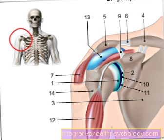
Vertebral artery
The Vertebral artery has its origin in the Subclavian arterywhich runs from the middle of the body to the shoulder behind the collarbone. The Vertebral artery then runs in pairs on the cervical spine as Arteria vertebralis dextra (right) and Left vertebral artery (Left). Here it runs in the Foramina transversariawhich can be described as small holes on the transverse processes of the vertebral bodies. Here it can Osteophytes (Bony outgrowths) that can lead to fainting due to pinching of the arteries.
Then the two step through Vertebral arteries along with the spinal cord that Foramen magnum, a large opening at the bottom of the skull bone. The Anterior spinal artery submitted. This supplies the spinal cord. In addition, the Inferior posterior cerebellar artery (PICA), submitted. This supplies the cerebellum. The two branches of the Vertebral artery to Basilar artery, which arterially supplies the brain stem via several small branches.
More information can be found here: Vertebral artery
Femoral artery
The Femoral artery (Femoral artery) is the largest artery on the thigh. It is a continuation of the External iliac artery below the bar. At this point, below the inguinal ligament, you can also check the pulse of the Femoral artery Keys. In addition, this section is often used for arterial access, for example for a contrast agent display of the coronary vessels. As important departures of the Femoral artery are the arteries epigastrica superficialis, A. circumflexa ilium superficialis, A. profunda femoris (which, as a strong side branch, supplies large parts of the thigh and hip), Aa. External pudendae (usually two) and A. descendens genus to call. The orientates itself in its course Femoral artery then on Sartorius muscle, which acts as the main muscle. The artery then runs inside the adductor canal (the canal between the adductor muscles) on the inside of the thigh. There it steps just above the hollow of the knee, on Hiatus adductorius (Adductor slot). Finally, it is called Popliteal artery (Popliteal artery) continued.
More information can be found here: Femoral artery
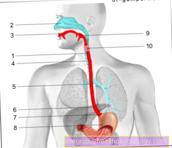
Carotid artery
The carotid artery is called the Common carotid artery, which runs as a strong artery on both sides of the neck and is decisive for the arterial supply of the neck and head responsible is. It arises directly from the aortic arch on the left side. On the right, it arises from the Truncus brachiocephalicus. The Common carotid artery within the Carotid vagina, a covering made of connective tissue. You can easily determine the pulse here on the artery if you feel at the level of the larynx and to the side of it. Because of this, the Common carotid artery also known colloquially as the carotid artery. The Common carotid artery then divides into two further arteries, the External carotid artery and Internal carotid artery.
At the junction you will find the so-called Carotid bodywhich registers the oxygen and carbon dioxide pressure in the blood. Besides, he takes that PH value, thus the degree of acidification of the blood is true. In addition, is due to the branching of the Common carotid artery of the Carotid sinus, which registers the blood pressure. With the information collected here, the body can react to fluctuations and regulate the various parameters. In the end there is External carotid artery several branches to the face, larynx, throat and thyroid gland. The Internal carotid artery pulls into the cranial bone and is involved in the arterial supply of the eye and brain. Because of this, a Stenosis (Narrowing) of the carotid artery or Internal carotid artery very risky. If the flow of blood is too low, the brain is undersupplied. If there is a narrowing on one side only, it can usually be compensated for from the other side.
This article might also interest you: Carotid artery
Pulse artery
The pulse artery is called in technical terms Radial artery referred to as they were on radius (Spoke) running along. The Radial artery originates from Brachial artery (Upper arm artery). It then runs down the inside of the forearm where the thumb is pointing. The Radial artery in its course is based not only on the spoke but also on Brachioradialis muscle. You can see this when you bend your hand towards your thumb. They are called Radial artery Pulse artery, as you can optimally get the right in front of the wrist Can feel the pulse. Here you go approx. 3 cm from the underside of the ball of the thumb down the inside of the forearm and feel with your index fingers between the central tendons and the lateral bone.
Just before the wrist is there Radial artery the Ramus palmaris superficialis (Superficial palm arch). This is a small artery that connects with the Ulnar artery combines and thus supplies the palm. The rest of the Radial artery pulls in front of the ball of the thumb on the back of the hand and supplies the thumb and one side of the index finger with oxygen-rich blood. Then ends Radial artery in the so-called Arcus palmaris profundus (deep palm arch), which is also with the Ulnar artery shorts. Thus the arterial supply of the hand takes place from two sides and is thus secured.
More information can be found here: Pulse artery
Coronary arteries
The coronary arteries, also called the coronary arteries or Coronary artery (Lat. Coronarius "crown-shaped") are the so-called "Vasa privata" (own vessels) of the heart. They serve exclusively arterial supply of the heart and are therefore of immense importance. Here they pull from the outside of the muscle as small branches into the muscle. A distinction is made between two coronary arteries, the Left coronary artery (left coronary artery) and the Coronary artery dextra (right coronary artery). They are branches of the ascending part of the aorta, i.e. branch off immediately behind the exit of the heart.
The Left coronary artery divides into one Ramus interventricularis anterior (RIVA) and one Ramus circumflexus (RCX). Of the Ramus interventricularis anterior runs as a branch of the left coronary artery to the apex of the heart. Of the Ramus circumflexus pulls down the left side of the heart and supplies its underside. The anatomy here often varies from person to person, but the course described applies to approx. 75% of the cases. The right coronary artery curves back and down on the right side of the heart. Here she takes care of e.g. the right atrium, the sinus node and the AV node, the clocks of the heartbeat. If the right coronary artery is blocked, there is an acute danger to life, as the heart no longer receives any impulses and therefore no longer beats.
More information can be found here : Coronary arteries
Popliteal artery
The Popliteal artery (Lat. Poples "hollow of the knee") is the continuation of the Femoral artery (Femoral artery). It begins with the exit of the Femoral artery from the Hiatus adductorius (Adductor slit) above the hollow of the knee. First, at the top of the knee, the Arteria superior medialis genus (Upper central knee artery) and the Arteria superior leteralis genus (Upper lateral knee artery) delivered to the inside and outside of the knee. Then the Popliteal artery into the hollow of the knee. The artery's pulse can be felt well here, as it is hardly covered by other structures. On the other hand, the artery here is also very prone to injury, which can lead to high blood loss. There is in the hollow of the knee Popliteal artery the Arteria media genus (Middle knee artery), which supplies the cruciate ligaments. Then the Popliteal artery further in the direction of the lower leg from the hollow of the knee and gives off two further branches that Sural arteries (Lat. Sura "Wade"). These supply the Gastrocnemius muscle, a two-headed, strong calf muscle. Finally, below the hollow of the knee, it divides Popliteal artery in the Anterial tibial arteryr and Posterior tibial artery on.
Gas and mass transfer
The exchange of substances between the blood and the environment takes place in the capillaries. This is favored by the very thin vessel wall and the huge total surface area of all capillaries. Some substances, such as gases, can cross the vessel wall unhindered, while other substances, on the other hand, are made via special ones Transport mechanisms absorbed into the tissue. The permeability of the vascular wall varies greatly from organ to organ. A continuous vessel wall (Endothelium) has a different degree of permeability depending on the organ (permeability). A fenestrated vessel wall (Endothelium) mainly lets water molecules through, while a discontinuous vessel wall (Endothelium) is completely permeable to all components of the blood.
arteriosclerosis

arteriosclerosis is the collective term for all pathological changes in arteries. These changes can have different causes. The most common form is Atherosclerosiswhich in our part of the world is often equated with arteriosclerosis. This pathological change is often found in large and medium-sized vessels and is favored by damage to the innermost vascular layer. Due to this damage, the smooth surface of the artery is roughened and components of the blood such as cholesterol, phagocytes (Macrophages) and fats accumulate and form a larger plug (atheromatous plaques) develop.
This results in a narrowing of the vascular space (Stenosis) and possibly to a reduced blood supply to the tissue behind it. If an artery closes as a result of a very large plug, the tissue behind it dies off, as it can no longer be supplied with oxygen and nutrients. This is called a heart attack.
These vascular changes are normal with age, but can be caused by various risk factors such as Smoke (Nicotine abuse), high blood pressure or Diabetes (Diabetes) are extremely favored.
PAOD
The PAVK, short form of Peripheral arterial disease, is a disease of the arteries. Here it comes to a Stenosis (Narrowing) or occlusion of arteries, in most cases due to arteriosclerosis. Count as a risk Diabetes mellitus (Diabetes), smoking, high blood pressure and lipid metabolism disorders, i.e. too high levels of fatty acids and cholesterol in the blood. The legs are often affected, which then hurt due to insufficient arterial supply. The result is that you can only walk short distances, which is why the PAVK was also nicknamed "intermittent claudication". The examination of the skin color (side by side) can be viewed as a simple diagnosis.If the skin on the foot is very pale and cold compared to the opposite side, there is most likely a circulatory disorder.
However, there are many more specific research methods. The symptoms can vary, depending on the degree of closure. In stage I, those affected do not notice anything about their illness. In stage II one subdivides between IIa, whereby the affected over 200 metres can walk continuously, and IIb, whereby those affected can walk continuously below 200 meters. In stage III pain occurs at rest. In stage IV it comes to necrosis (Tissue death). A distinction is also made here between IVa and IVb. Stage IVa results in dry necrosis due to insufficient blood flow. The fabric turns black. Stage IVb results in a bacterial infection of the necrosis. The problem here is that the bacterial infection is difficult to fight because the body's immune defenses cannot be transported to the infection via the undersupply. The therapy of PAOD ranges from Lifestyle, drugs, bypass operations and amputation of the dead tissue.
Further information can be found here: Peripheral arterial disease