pool
English: Pelvis
Medical: Pelvis
anatomy
The basin (Pelvis) refers to the part of the body above the legs and below the abdomen. In humans, a distinction is made anatomically between a large (Pelvis major) and a small basin (Pelvis minor).
The urinary bladder, rectum and genital organs are located in the pelvic cavity, in women the uterus, vagina and fallopian tubes, in men the prostate. At the same time, the term "pelvis" is also used for the bones in this region of the body, which is made up of the two hip bones (Ossa coxae) and together with the sacrum (Sacrum) forms the so-called pelvic belt or pelvic ring. The pelvic ring as part of the human skeleton connects the spine via the sacrum-iliac joint (Sacroiliac joint) and the hip joint with the lower extremities. Thanks to its stability, the pelvis gives people a secure stance and an upright posture.
Illustration of the pelvic bone

Pelvic bones
(Pool - Pelvis)
- Third lumbar vertebrae -
Vertebra lumbalis III - Iliac crest - Iliac crest
- Iliac scoop -
Ala ossis ilii - Sacrum-iliac joint
(Sacroiliac Joint - SI joint)
Articulatio sacroiliaca - Sacrum - Sacrum
- Tailbone - Os coccygis
- Ischium - Os ischii
- Pubic bone - Pubis
- Femur - Femur
- Pubic symphysis -
Pubic symphysis - Lumbar cruciate ligament kink -
Promontory
A - pelvis from the front
B - from the outside
You can find an overview of all Dr-Gumpert images at: medical illustrations
Hip bone
Anatomically, that's in the pelvis right and left hip bone, each made up of three parts, the Iliac bone (Os ilium), the Pubic bone (Pubis) and the Ischium (Os ischii). These three bones are used in child development created separately from each otherso that the pelvis can grow symmetrically with the body and merge around the age of 15 in the area of the acetabulum (Acetabulum) together to form the now uniform hip bone. The acetabulum forms with the head of the Thighbone (Femur) the hip joint. Since this joint is exposed to heavy loads throughout its life and wears out over the years, older people often experience complaints there. The Sacrum and iliac joint connects the hip bones with the sacrum. This joint is very tight and can hardly make any movements, but it plays an important role in the Suspension of the spine. Due to incorrect posture or injuries, the joint surfaces in the sacrum and iliac joint can shift against each other, which causes severe pain (so-called ISG syndrome).
Also read: ISG pain
At the front of the bony pelvis, the hip bones connect via a cartilaginous connection, the Pubic symphysis (Pubic symphysis).
This connection point plays in the pregnancy an important role through the influence of Hormones the pubic symphysis becomes softer and more flexible, so it fits with the birth the child's head better through the pelvis.
The female and male bony pelvis differ in their structure. The two are with the woman Cantilever bladeswhile the male Pelvis high, narrow and tight is created. The female pelvic entrance is larger and roundish oval, the male, on the other hand, is more heart-shaped. The pelvic outlet is also wider in the female pelvis.
Symphysis
The term symphysis has two meanings in medicine.
In itself one understands by the term Symphysis the connection of bones through Fiber cartilage. The symphysis therefore falls under the category of "fake joints", since it is real Joints a Bone gap is found between the bones and has no cartilaginous connection. False joints, in turn, are divided into synchondroses and symphyses. Synchondroses contain hyaline cartilage, symphyses contain fiber cartilage. Among other things, symphyses can be found in the body in:
- the Pubic symphysis (Called symphysis pubica)
- in the Intervertebral disc
- in the Lower jaw and
- in the Sternum
As the changes in the Pubic symphysis plays an important role in pregnancy, the pubic symphysis is often referred to by medical professionals as the symphysis instead of the pubic symphysis. Therefore, when you hear the word “symphysis” in everyday medical practice, you tend to think of the Pubic symphysis than the bone connection.
The pelvic symphysis is located between the right and left pelvic bones. The fact that this area is not properly ossified plays a very important role in childbirth. Because the fiber cartilage connection allows the pelvis to expand easily.
Due to the hormonal changes during pregnancy, the fiber connection changes and thus becomes more flexible.
The So the pelvis can widen and thus creates a larger birth canal. However, stretching the pelvis can be painful even before birth. If the stretching becomes too strong, it can lead to Symphysis loosening come which can also cause severe pain.
It can also be used in birth Rupture (Rupture) the symphysis come, this must then be treated after the birth with training of the muscles and a so-called trochanter belt.
The pelvic symphysis also forms the starting point for the straight line Abdominal muscle, the so-called rectus abdominis muscle. It pulls from the ribs, more precisely from the fifth to the seventh rib down to the pubic bone and is in the trained state as "Washboard abs" known.
Pelvic inclination
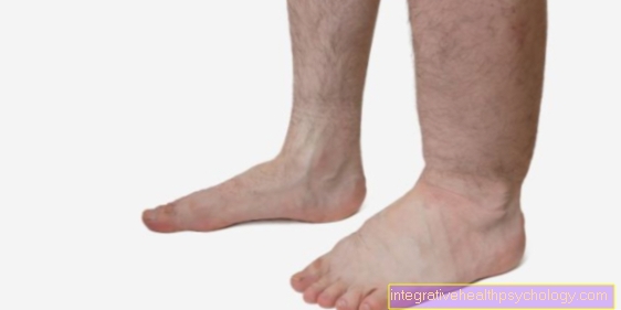
Common cause of Back pain is a Malposition of the pelvis. By legs of different lengths For example, the pelvis can be inclined, which does not necessarily lead to discomfort, as the body can compensate for many inaccuracies. Is the Pelvic inclination but seriously, it threatens in the long run lateral curvature of the spine (Scoliosis), which is common too Muscle tension, Joint wear and complaints in everyday life.
In medicine, one differentiates pelvic obliquity into two different categories.
- A functional misalignment is mostly through muscular tension the lower Back and gluteal muscles caused by one-sided loads, for example at Sports can be triggered. For example, if the tension is relieved with the help of physiotherapy solve, the functional pelvic inclination is also eliminated again.
- A structural misalignment of the pelvis is against it anatomically caused and does not return to a straight position. The causes of the structural pelvic inclination are Operations, accidents, prostheses or genetic predispositions. In most cases, legs of different lengths are the reason for the pelvic inclination. About one Leg length difference of six millimeters and more discomfort can arise. Then orthopedic advice should be obtained, usually in such cases an compensating shoe insert used.
Pelvic pain
The term Pelvic pain can indicate different types of pain and their localization. Since there are other terms that can describe a more precise location of the pain, these terms should be used for the exact localization of the pain if the symptoms are present.
When it comes to pain the Bone structures or Muscles in this area, one would think of Hip pain speak. However, should be a organ Cause pain, which is in the pelvic area, other pain can occur. In this case you would go by stomach pain, or more precisely from Abdominal pain or for example Bladder pain speak. Depending on whether the pain is felt when moving the hip or occur at rest, or how intense the pain is, these terms can clarify the affected region of the complaints.
Pain in the pelvic bone becomes common
- from injuries like Bruises or breaks
- Pain from the Decrease in bone tissue (osteoporosis) or
- by Posture faults triggered.
Pain in the uterus, the Ovaries, the prostate or the bladder have various causes. A Appendix irritation or a tumor can trigger such pelvic pain, for example. Abdominal inflammation in the pelvic area, usually the result of Genital infections, often by pathogens such as bacteria, Viruses or fungi are also associated with significant pelvic pain. Often a psychosomatic pain occurrence come into question for pain that radiates into the abdomen and pelvic area. But also Diet errors, chronic bowel disease, food intolerance, ectopic pregnancy, or infection can cause pelvic pain.
The hormone-related loosening and the pressure-related stretching of the Pubic symphysis in the pregnancy can also cause pain in this area. Up to ten percent of all pregnant women are affected by such complaints, which are particularly pronounced when standing and walking. In rare cases, childbirth can lead to a full one Division of the pubic symphysis come (symphysis rupture).
In any case, it is advisable for pelvic pain to see a doctorto find out the cause of the pain. Therapy then depends on the diagnosis.
Causes pelvic pain
The causes of pain that occur in the pelvic area can be very different. So can Joints or Muscles the hip as well as the Back Cause pain, which is then called Pelvic pain be perceived. Also one Misalignment of the hipscaused, for example, by legs of unequal length, can cause pain. A present osteoporosis or injuries in the pelvic area often cause typical pain in that area. Other pain can arise when organs in the pelvic area are affected. So diseases of the bladder, prostate or the female genital organs Cause pain when inflamed or affected by a medical condition. Also diseases of the Intestines trigger pain, which can then radiate into the pelvis. Radiated pain can be from a Appendicitis or one Diverticulitis be evoked. Also pain that is related to the diet, for example in Food intolerance can cause problems in the pelvic area.
Diagnosis of pelvic pain
At unspecific pain of the pelvis, a doctor should be consulted in any case. This is particularly important if the reason for the symptoms is unclear and the pain is of great intensity.
In many cases, a diagnosis can be made through a detailed doctor-patient conversation and a physical examination.
Imaging procedures such as Ultrasonic and especially that MRI of the pelvis can simplify the diagnosis. Depending on the intensity of the pain, an immediate referral to a clinic should be made so that serious illnesses that require immediate surgical treatment can be excluded if necessary.
Further information can also be found under our topic: MRI of the pelvis
Injuries and diseases of the pelvis
In the area of the bony pelvic girdle there are often joint diseases. For example, it can be a Joint wear (arthrosis) come. Also Inflammation of the joints (so-called Coxitis) often come in the area of the Hip joint in front. The cause of such a joint inflammation can be varied. For example, you can by bacteria which may have entered through open wounds or operations, or through a Inflammation of the bone marrow (Osteomyelitis) that has penetrated the hip joint. But also as a result of rheumatic diseases (Rheumatoid arthritis) or symptomatic as a side effect of an underlying disorder such as a Coxarthrosis hip joint inflammation can occur.
The so-called Sacroiliac joint syndrome (ISG syndrome) is one painful tilting of the articular surfaces of Sacrum and iliac joint into each other and is a common cause of back pain. Other pelvic diseases are the reduction in bone tissue (osteoporosis), Inflammation of the bones (Osteomyelitis) or also Bone tumors.
The pelvic belt is very stable and robust and looks like a ring when viewed from above. Because of this stability will be Pelvic fractures often only due to serious accidents caused as it happens in traffic or when falling from a great height. In people who suffer from reduced bone density, even lighter falls can lead to pelvic fractures. A distinction is made depending on which area of the pelvis is affected Fractures of the pelvic ring of Fractures in the area of the acetabulum. The therapy for these pelvic fractures depends on the stability of the fracture and can either conservative or operative respectively.
Also read: SI joint osteoarthritis
Pelvic diseases in children
Children or teenagers at the age of 3-10 years often suffer from a so-called Runny nose (Coxitis fugax). This denotes a germ-free inflammation of the hip jointthat result in sudden onset in the knee radiating pain and one Restriction of rotation of the leg in the hip joint. The common cold usually disappears spontaneously within one to two weeks, and the disease is often one viral cold ahead. By a Ultrasound examination the diagnosis is confirmed and rest is usually sufficient to cure the hip runny nose.
Under Perthes disease one understands an orthopedic childhood illness, in which by circulatory disorders Bone tissue in the femoral head dies. Perthes' disease manifests itself through limping, knee pain and restricted movement in the hip area.
pelvic floor
The pelvic floor forms the lower limit of the pelvic canal and is a connective tissue-muscular structure.
The functions of the pelvic floor include Securing the position of the abdominal and pelvic organs and the support of the closure of the anus (anus) and urethra (urethra). This is done by tensing, relaxing and reflexively countering the Pelvic floor muscles reached. Tensioning is used to secure continence, the sphincter muscles of the urinary bladder and anus are responsible for this and are largely supported by the pelvic floor muscles. The pelvic floor relaxes during urination, defecation and sexual intercourse; during orgasm it pulsates, which means that tension and relaxation alternate. The reflective countermeasures is important, for example, when coughing, sneezing and laughing, as otherwise the increase in pressure in the abdomen can lead to urine loss. The human pelvic floor is through Obesity, poor posture and operations in the small pelvis weakened, in women additionally by Births. This can be too lack of control of the excretory organs, Vaginal or rectal prolapse, bladder and uterus subsidence. In many cases this weakness can get through Training the pelvic floor muscles be fixed again.


.jpg)