Pelvic bones
General
The bony pelvis (pelvic bone) is made up of the two hip bones (Os coxae), the tailbone (Os coccygis) and the sacrum (Sacrum) together.
It is used for the articulated connection of the spine with the lower extremity. In addition, the bony structure differs between the sexes due to the anatomical requirements for the birth of a child.

function
The pool mainly serves as the articulated connection between the Spine and the lower extremities.
It is especially true with the spine articulatedbut very firm connectedso that hardly any movement is possible here. However, it thereby enables a safe was standing and an upright one attitude.
In addition, the bones are approach- and Points of origin for numerous Muscles.
construction
The bony pelvis consists of the
- Sacrum (Sacrum), the
- coccyx (Os coccygis) and the
- both hip bones (Os coxae dextrum et sinistrum).
A hip bone, in turn, is made up of three different bones:
- Os ilium (Iliac bone),
- Os ischii (Ischium) and
- Pubis (Pubic bone).
The pelvis will be roughly in a great and a little one Pelvis articulated. The Terminal line to separate these two parts of the pelvis. This is an imaginary dividing line at the most prominent point of the 5. Lumbar vertebra begins and goes from there to the symphysis.
The space between the two iliac blades above the linea terminalis is called the large pool (Pelvis major), the space below it as small basin (Pelvis minor). The small pelvis narrows downwards and thus represents the actual one Basin funnel The individual parts of the pool are to be explained in more detail below.
Illustration of the pelvic bone

Pelvic bones
(Pool - Pelvis)
- Third lumbar vertebrae -
Vertebra lumbalis III - Iliac crest - Iliac crest
- Iliac scoop -
Ala ossis ilii - Sacrum-iliac joint
(Sacroiliac Joint - SI joint)
Articulatio sacroiliaca - Sacrum - Sacrum
- Tailbone - Os coccygis
- Ischium - Os ischii
- Pubic bone - Pubis
- Femur - Femur
- Pubic symphysis -
Pubic symphysis - Lumbar cruciate ligament kink -
Promontory
A - pelvis from the front
B - from the outside
You can find an overview of all Dr-Gumpert images at: medical illustrations
Hip bone (os coxae)
The hip bone consists of three parts, which fuse together, creating a hip bone.
The Y-shaped fusion joint the part is in the Acetabulum (Acetabulum). The two sides of the hip bone are over the Symphysis (Pubic symphysis) and the Sacrum (Sacrum) connected to a bone ring.
The two hip bones are each over the Sacrum- Iliac bone- joint (Articulatio sacroiliaca) articulated to the sacrum. This is a Amphiarthrosis, i.e. the two bones are very tightly connected and hardly allow any freedom of movement. However, the joint is used to suspension the spine is very important.
The Iliac bone (Os ilium) takes up most of the hipbone and can be expanded into a wide expanse
- Iliac scoop (Ala ossis ilii) and the
- Iliac body (Corpus ossis ilii) structure.
The border of these two parts forms a bone-shaped bar that Arcuate line. At the same time, this line also represents the border between the large and small pelvis.
There is a small one on the inside of the iliac blade pit, the Iliac fossa. This serves as the origin of the Iliacus muscle. The outside is called Gluteal facies designated. It has three bone lines that serve as attachment points for the gluteal muscles.
The Iliac scoop has an upper thickened edge, which as Iliac crest (Iliac crest) referred to as. This runs towards the front in the Anterior superior iliac spine, to the back of the Posterior superior iliac spine out. In each case there is another bone protrusion, which is accordingly called Anterior inferior iliac spine and Posterior inferior iliac spine referred to as.
The Ischium (Os ischii) is also converted into a
- body (Corpus ossis ischii) and one
- Edge part (Ramus ossis ischii) structured.
The corpus forms the largest part of the Acetabulum (Acetabulum) and runs backwards in the so-called Ischial spine (Sciatic spine) out. This separates the large and small bony incision of the Ischium (Major ischial notch and minor).
Below the smaller notch is the Ischial tuberosity (Sciatic tuberosity), which is the origin of the hamstring muscles. That too Pubic bone (Pubis) is divided into three parts:
- the Pubic body (Corpus ossis pubis) and the
- upper and lower Edge of the pubic bone (Ramus superior and inferior).
The pubic bones on both sides protrude over the Symphysis in the bony connection and thereby form the pelvic ring. There is a lateral to the symphysis Bony prominence, the Pubic tuberosity. A bone ridge pulls from there Symphysis (Crista pubica), another for Acetabulum (Obturator crest).
Together with the Ischium (Os ischii) surrounds the pubic bone hole in the pool (Obturator foramen).This hole is through a membrane (Obturator membrane) closed so that only the Obturator nerve can pass through. This membrane is used for Obturator muscle internus and externus as the origin.
The Acetabulum (Acetabulum) is formed by all three bone parts and appears as a bony, circular depression that is surrounded by a bony bulge. The acetabulum is crescent-shaped with cartilage covered and provides the articulated connection to the femoral head of the Thighbone represent.
Sacrum
The Sacrum is merged by the five Sacral vertebrae and the ossified ones in between Intervertebral discs educated.
It will be a
- lying in front concave Area, from one to
- rear lying convex Differentiated area.
The one lying down top (caudal) of Sacrum is called Apes ossis sacri, the most prominent point at the base of the sacrum as Promontory designated.
The Sacrum canal (Sacral canal) represents the continuation of the Vertebral canal represent.
That too Sacrum carries different surfaces, which as Articulated joints act to neighboring structures. The two are lying to one side Facies auriculariswhich corresponds to the area of the Ilium (Os ilium) forms an articulated connection. In addition, the Sacrum small holes between the vertebrae (Intervertebral foramina), which is the exit point of the Spinal nerves represent.
Coccyx (coccyx)
The coccyx adjoins the sacrum and usually consists of four Vertebral bodies. However, the number can vary.
The individual vertebrae are through Synchondroses connected so here no movements can take place. The size of the individual vertebrae decreases towards the bottom.
Basin dimensions
Be at the pool outer and inner Differentiated basin dimensions.
In addition, cross and inclined diameters certainly.
- The smallest sagittal diameter between the Back surface the symphysis and the Promontory is approx. 11cm.
- The greatest transverse diameter in the course of the Terminal line is on average 13.5cm.
- The distance between the Lower margin the symphysis and the promontory 12.5cm.
These measurements should be especially important during a pregnancy be determined in order to be able to give an assessment of whether the child during birth problem-free the bony pelvis can pass.
Gender differences
The basin has typical sexual Differences on.
So is the basin entrance of the Man rather card heart shaped, that the Mrs. queroval.
In addition, the Pubic angle with the man acute-angled (approx. 70 °) more likely for women obtuse angled (approx. 100 °).
The iliac scoop itself is more likely to turn up in a man steep it is more to the side for women expansive and the shape of the pelvic ring is on a man high, narrow and closelywhile she is at the woman low, wide and far is. All of these differences serve the optimal ones requirements for one birth.
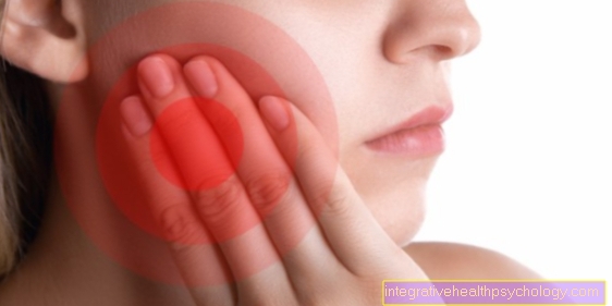
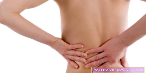
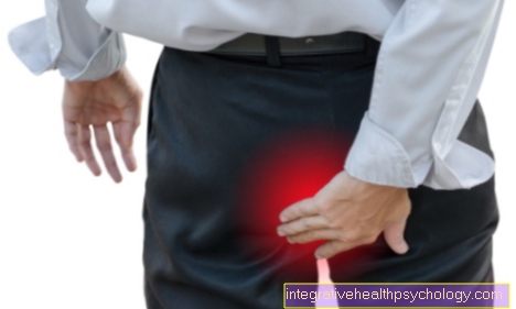
Pelvic bone pain
Pelvic pain usually affects that hip joint, the lower back, the groin, or the ligaments and muscles that run around the hips. Only in rare cases can the pain actually be traced back to the pelvic bone. Often there are shifts from Sacroiliac joint (short ISG) that can arise from different leg lengths, back pain, muscle defects or twisting of the bones in the joint.
Tension, irritation of the ligaments or tendons, a pinched nerve or defects or injuries to the internal organs can all be the cause of pain in the pelvic bone. Furthermore, fractures, sprains or strains can also be the reason. The pain often radiates to the leg, back or groin. If the pain does not go away after a few weeks, a doctor be sought out.
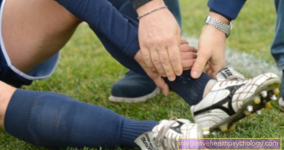
What to do in case of an ISG blockage?
Should the pelvic bone or the Sacroiliac Joint (ISG) shifted and thus the mobility of the joint be restricted, one speaks of a ISG blockage. This usually manifests itself in pulling pains, which intensify as soon as the leg is in the hip rotates outwards (e.g. sitting cross-legged or crossing your legs). If this is the case, a lot of heat should be applied to the painful area (e.g. using a hot water bottle) so that the muscles around the joint can relax. Often times, careful and correct exercise at home can relieve the pain.
One exercise consists of lying on your back on the floor. The legs are put down at an angle. Now you can swing carefully from left to right while your feet are constantly touching the ground. Another exercise is the so-called. Cat hump. You stand on four feet and carefully hunch your back. Then let it sag slowly. If the complaints cannot be resolved independently, a osteopath or a Physiotherapist be sought out. This can then determine the exact cause of the ISG blockage and rectify it. In order to prevent injuries of this kind, regular and stabilizing exercises for the core and pelvic muscles must be completed.
You might also be interested in the following articles: Dissolve an SI joint blockage, Therapy of an ISG blockade
Summary
The bony one pool is made up of three different bones, which penetrate each other fixed joints are connected to each other.
These bones include that
- Hip bone, the
- coccyx and the
- Sacrum.
The basin serves on the one hand connection between Spine and lower extremity and the stabilization, as well as the upright gait.
There are also some organs in the basin and it is Penetration point of the child during a birth. For this reason, there are numerous differences between the basins in the structure and shape of the basin Genders.