Importance of the biopsy for breast cancer diagnosis
Synonyms in a broader sense
Biopsy, fine needle aspiration, punch biopsy, vacuum biopsy, MIBB = minimally invasive breast biopsy, excisional biopsy

Biopsy (tissue sample)
Often, despite exhausting all diagnostic possibilities, only a biopsy finally provides clarity as to whether it is one benign or one malignant tumor acts. So if a biopsy is done, it doesn't necessarily mean you have cancer. It is possible today to see almost all abnormal or suspicious findings in the chest to biopsy, i.e. take a sample and make a diagnosis. A biopsy is easy to perform, hardly puts any strain on the breast tissue and can usually be done without hospitalization, but the examination can be painful. The sample is then taken by a pathologist - a Specialists in tissue and cell studies - examined. The pathologist can use the cells in the tissue to make his diagnosis, as cancer cells look different from healthy cells. One speaks of a tissue or histological examination.
Read more about this topic here: biopsy
In the past, an incision had to be made to remove a piece of tissue. So-called minimally invasive procedures are used today, in which samples are taken with needles in order to protect the breast tissue as well as possible.
There are numerous methods of doing this, from extremely thin needles to relatively thick hollow needles. The thought of being stuck in the chest with a needle is terrifying for most women. The most uncomfortable part of the examination is the moment when the skin is pierced. Depending on the diameter of the cannula used, you will feel less or more pain, comparable to a blood sample. The skin is first anesthetized locally. The actual movement of the needle in the breast tissue, however, can hardly be felt.
The biopsy option can prevent many unnecessary operations (see also breast cancer surgery). The different methods can basically be divided into two categories. In the case of palpable findings, fine needle puncture and ultrasound-guided punch biopsy are possible methods. In the case of findings that can only be detected by mammography, stereotactic biopsy procedures are possible (see below).
If the findings are recognized as benign after the tissue sample has been taken, no further intervention is required. The further procedure depends on the patient's complaints. The lump can be removed if it causes pain, continues to grow, or is simply bothersome and / or unsettling.
Depending on the size of the lump, however, this can result in retractions, changes in shape and scars on the breast, which in turn can lead to pain.
Read more on the subject at: Breast biopsy
What can the pathologist recognize from a tissue sample?
Based on the tissue sample, the pathologist can first provide information on whether the change is benign or malignant. A more positive Finding means in this context that the finding is positive for cancer, i.e. malignant. The other way round is with one negative Finding no evidence of cancer. In other examinations, too, "positive" always means in the language of pathologists that something has been detected or is present, not that it is a "good" result for the patient.
The pathologist can also determine the origin of the cells. This means that he can generally tell whether a lump in the liver is liver cancer or whether, for example, the daughter tumor of a breast cancer is present. The pathologist uses the tissue sample to create a kind of “tumor profile”, ie a list of the characteristics of the tumor. The treating physicians can use this as a basis for their therapeutic approach and make statements about the prognosis of breast cancer.
If there are changes in the cells, the pathologist creates a "grading". The grading of the cells indicates how much the cells still resemble their original tissue or, conversely, how much they have changed. This is also called the degree of differentiation of the cells. In addition, characteristic changes in the cell nuclei and the occurrence of necrosis (dead tissue) respected. The “grading” of the cells influences the prognosis and the possible treatment strategies and indicates the aggressiveness of the tumor.
Please also read our page Breast cancer stages
Using various test methods, the pathologist can also make statements about other properties of the cells that make them particularly sensitive to certain forms of therapy and at the same time have an impact on the prognosis. This includes certain receptors that some tumor cells have and some do not.
Please also read our page Tumor markers in breast cancer
Info: Degree of differentiation
The higher the grading, the less the tumor cells resemble healthy cells and the more aggressive the cancer.
There are three levels:
• G1 means "well differentiated",
• G2 means "moderately differentiated",
• G3 means "poorly differentiated".
About 60 out of 100 tumors are classified as G2.
Examination of the tissue sample
A biochemical examination is carried out on the tissue sample to determine the sensitivity and quantity of the hormone receptors on the cancer cells. H. the amount of receptors for the female sex hormones estrogen and progesterone. Since tumor cells are characterized by the fact that the normal functions of a cell are disturbed, it can also lead to a loss of the ability to produce receptors for the sex hormones. A distinction is made between estrogen receptor positive and negative breast cancer cells (types of breast cancer). This distinction plays a role above all in terms of therapy options. If there are many receptors, it is a sign that the cancer is responding well to hormone therapy.
Another examination of the tissue sample will determine whether the cells of the tumor contain many HER2 / neu receptors. Via these receptors, growth factors can “dock” to the breast cancer cell and stimulate it to divide more and the tumor to grow faster. This docking can be prevented with antibody therapy.
Read more on the topic: How do you recognize breast cancer?
What are the risks of a biopsy?
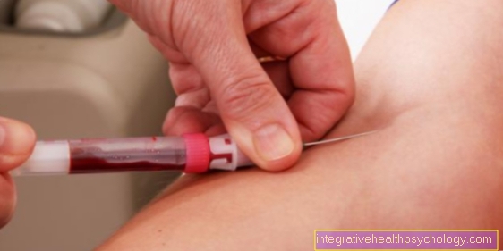
With everyone biopsy there is a small risk of infection and / or bleeding. Can through the puncture channel bacteria penetrate the breast tissue and cause inflammation there, but this is extremely rare. Through the puncture in the chest Blood vessels be injured, which in turn leads to smaller ones Bleeding can lead. The only danger here is if the patient e.g. has a blood clotting disorder or anticoagulant drugs occupies (e.g. aspirin). In order to clarify this in advance, blood is taken before each biopsy and the coagulation of the blood is examined, as well as a list of those taken Medication created.
Read more about the topic here: biopsy
How long does the sampling take?
How long it takes to take a tissue sample in each individual case depends on both the type of biopsy and the doctor performing the test. It is mostly done in an outpatient setting, which means that patients can usually go home after the rehearsal. The removal normally takes a few minutes and is checked using imaging techniques. The puncture of the needle is barely noticeable, as the skin and the tissue in the puncture area are temporarily made insensitive to pain with a local anesthetic. After collection, a pressure bandage is placed over the area of the tissue sample. To avoid acute complications, the patients should remain under medical care for a few hours to avoid secondary bleeding, but they can return home on the same day.
What should be considered after removal?
The biopsy to classify a benign or malignant finding in the breast is a frequently performed procedure and rarely leads to complications. A slight bleeding and thus the formation of a bruise in the area of the tissue sample is normal and disappears after a few days. Depending on the collection method, a small scar can form. A complication of this can be the formation of a scar growth, which usually only becomes a cosmetic problem. However, if the breast continues to bleed heavily and persistently in the days after the procedure, this may be an unwanted consequence of the biopsy and should be presented to your doctor immediately. In order to avoid this consequence, it is generally recommended to wear a tight-fitting pressure bandage and bra and to take care of yourself after taking the sample. In rare cases, the procedure can also get bacteria into the punctured area, which could potentially cause inflammation. It is therefore important not to go to swimming pools or saunas in the hours or days after the sample is taken. If the puncture site becomes red, swollen, overheated or becomes more sensitive to pressure, your doctor should be consulted to rule out inflammation or, if necessary, treat it in good time.
Are cancer cells carried over during a biopsy?
As this question is asked frequently, this risk should be given special attention. Patients often fear that taking a tissue sample could spread cancer cells into the breast. This fear is essentially unfounded. Research has shown that Growth of single cancer cells in the punctured tissue is extremely unlikely. There are, however, differences between different forms of cancer and between the different collection techniques. For the two types of cancer where Biopsies are most frequently performed in diagnostics, Breast cancer and Prostate cancer So far there is no evidence that tumor cells that have been carried over have led to the emergence of new cancer foci. In other forms of cancer, however, it can be more common, e.g. with certain types of Ovarian cancer. The risk can never be completely ruled out.
Which Biopsy form is used at the end can only be clarified in an individual discussion with the attending physician. The following information therefore only represents general background information. There are always new modifications to the sampling techniques described, which differ in details and we are constantly trying to improve the current techniques.
Fine needle puncture
In the Fine needle puncture individual cells or cell clusters are taken directly from the node with the help of a syringe and a very fine cannula (only 0.5 mm in diameter, which is thinner than a pin). The results of the examination are usually available on the same day. The quality of the fine needle puncture depends heavily on the experience of the examiner. In the case of malignant findings, the diagnosis is 96% certain. With benign findings unfortunately only 90%, i.e. With a palpable lump, you cannot always rely on a negative result. Since only individual cells are removed during a fine needle aspiration and not entire pieces of tissue, it can be difficult for the pathologist to make statements e.g. about grading or type of growth to make. If necessary, a Punch biopsy be performed. The fine needle puncture is only used by a few specialized examiners and is increasingly being replaced by the punch biopsy.
Punch biopsy
The punch biopsy is another way of taking a tissue sample from an abnormal tactile and / or mammography finding. A needle with a diameter of approx. 1.6 mm is shot into the tissue at high speed. This technique ensures that inserting the needle is actually no more uncomfortable than taking blood. However, a small skin incision under local anesthesia is also necessary. An experienced examiner shoots the needle directly into the findings in question "under sight". Under vision means that an ultrasound of the breast is performed at the same time, on which the findings, the needle and its position can be seen. Usually three different punches are taken from three different areas of the tumor, but additional punches may also be required.
A punch biopsy can remove more tissue than a fine needle aspiration. Inside the needle is a cavity in which a tissue bandage can be received as a punch. The sample is then sent to the pathologist. With a punch biopsy, the diagnosis is almost as certain as surgical removal of the tumor; in the case of a malignant finding, the certainty of the diagnosis is 98%, and in the case of benign findings, the certainty is over 90%. The punch biopsy can avoid many unnecessary surgical interventions in the case of benign findings.
Stereotactic Procedures
The term stereotactic (stereo = spatial, taxis = order or orientation) procedures summarize various techniques in which work is carried out under X-ray control. By taking several images from different directions, the doctor can orientate himself spatially while performing the biopsy and localize the findings precisely.
Stereotactic procedures are mostly used for the biopsy of findings that are only used in the Mammography can be seen, e.g. with conspicuous Microcalcifications in the chest. The only difference between the various techniques is the needle used and the amount of tissue samples that are taken. Digital mammography is now mostly used as an X-ray control. In contrast to conventional mammography, the images are immediately available and the duration of the examination is thereby greatly reduced.
Stereotactic punch biopsy and fine needle aspiration
Both procedures run exactly as described above, with the difference that the Ultrasonic here through one Mammography machine is replaced. Taking the biopsy is somewhat more uncomfortable because the patient has to sit still for a long time while the breast is compressed in the mammography device for the recording. In addition, there is the radiation exposure from multiple recordings, which are necessary to localize the findings in three-dimensional space. Even with stereotactic punch biopsy / fine needle puncture, the reliability of the results is very high if the finding is made. However, only a few clinics have the technical capabilities for one stereotactic punch biopsy.
Vacuum biopsy (MIBB = minimally invasive breast biopsy)
The vacuum biopsy (MIBB = minimally invasive breast biopsy) is a further development of conventional minimally invasive needle biopsies. Another name for this method is mammotome vacuum biopsy. It is used when a mammography finds altered tissue five millimeters in size or more. The vacuum biopsy can be combined with both imaging methods, mammography and ultrasound. The combination with mammography is more common, which is why it is counted among the stereotactic procedures.
Most of the time, the patient lies on her stomach on a special examination table with an opening in which the breast is placed so that it cannot shift or slip during the examination. A hollow needle about three millimeters thick is used for the vacuum biopsy. After a local anesthetic, the hollow needle is inserted into the breast through a 3-4 mm long incision. By negative pressure (vacuum) tissue is sucked into the hollow needle, in which there is a tiny high-speed knife that separates the sucked in sample from the rest of the tissue. The tissue is then transported into an opening in the middle of the needle, from which it can be removed with tweezers. The needle can rotate around its own axis when the tissue is removed so that samples can be taken from several points of the findings and the environment. This increases the reliability of the diagnosis. Some clinics have special devices in which the vacuum biopsy can also be performed while sitting. In addition, with this technique, a microclip can be inserted after the samples have been taken, which marks the location of the sample collection for later control examinations or operations.
Excisional biopsy
A Excisional biopsy is a surgical procedure; it is therefore also called an operative or open biopsy. In general anesthetic the entire suspicious area is removed from the chest removed and then taken for examination by the pathologist. The final confirmation of the diagnosis can only be made by removing the entire breast lump with subsequent microscopic tissue examination respectively. Therefore, excisional biopsy is still the standard procedure in many centers. But it is also the procedure with the most side effects.
For many women, the 3-4 cm long scar that remains on the breast is very annoying, and tissue damage can also lead to retractions and adhesions within the breast. This makes it difficult to judge the following later Mammograms. Since most biopsies performed lead to negative results, some doctors ask themselves whether the benefit for women with a positive result compared to other less invasive procedures really outweighs the harm for women with a negative result.
Read more about this topic here: biopsy










.jpg)


