Radiation therapy planning
Note
This topic is a continuation of our page: Radiation Therapy
Synonyms
Radiation planning, planning of radiation therapy, preparation of radiation therapy
definition
Radiation therapy planning includes all preparatory measures that are necessary in order to be able to carry out radiation therapy according to the required quality criteria.
procedure
The following steps are usually carried out when planning treatment:
- storage
- Image acquisition
- Determination of the therapy region
- Calculate the treatment plan
- Simulation of the irradiation
- Transition to radiation therapy
storage
The positioning of the patient must be selected so that the patient can be reproduced comfortably and safely and that the treatment region can be reached as directly as possible by the therapy beam. The following are common examples:
- Storage during irradiation of breast cancer (cancer of the female breast):
Here the supine position with arms raised above the head or the oblique lateral position on the healthy side has prevailed. Various storage aids are used to avoid wobbling. Arm holders, wedge pillows, vacuum mattresses.
Through these measures, the chest is easily accessible to the therapy beam and the lungs, heart and chest on the other side can be largely spared. - Storage when irradiating prostate cancer (cancer of the prostate gland in men):
The patient lies on his back and a pillow is placed under his knees so that the pelvis can lie flat on the treatment table. A hollow back is avoided. The arms are on the chest. - Storage for radiation in the head or ear, nose and throat area:
Since the head is very mobile and at the same time critical regions are very close, the head must be fixed for radiation therapy in order to avoid damage to healthy organs (brain, eyes, nerves, etc.). Most often, head masks are made from thermoplastic material. This special plastic material is heated and thus stretchable. In this state it is pulled over the patient's head and attached to the treatment table. It adapts to the facial contour like a plaster cast. The material cools down in a few minutes and no further deformation is then possible. The mask is then put on before each treatment session.
Image acquisition / computed tomography

Once the positioning is complete, a computed tomography (slice x-ray) of the therapy region is made in this position. A three-dimensional model of the patient is calculated with this image data. Furthermore, with the computer tomography, the first target markings are drawn on the skin, which are required for the same positioning during the irradiation. There are three room lasers each in the room of the computer tomograph and in the treatment room of the linear accelerator. One is attached to the left, one to the right and the third above the treatment table. All three lasers meet at one point. In the accelerator room, this is the point that is exactly one meter away from the point in the accelerator head where the X-ray beam is generated. At the same time, the axis of rotation of the linear accelerator runs through this point, which it can bypass 360 °.
Most often this point is localized in the tumor after treatment planning has been completed.
If all three laser points coincide with the skin markings, the patient is in the same position during each irradiation as he was when planning.

Four slice x-rays at the height of the upper body
Determination of the therapy region
In the image data set obtained, the attending physician now marks which area should receive the therapeutic radiation dose and which areas and organs must be spared. Many aspects of the tumor disease must be taken into account. For some, it is sufficient to treat the tumor region yourself; for others, the lymphatic drainage must also be included, as metastases (settlements) in this region are more common in them. In addition, the radiation exposure of the healthy neighboring organs must be taken into account so that there is no permanent damage caused by radiation therapy.

Reconstruction of the pelvic organs in men:
Blue = rectum
Pink = prostate
Orange = seminal vesicles
Dark red = urinary bladder

Marked therapy region for prostate cancer (red circles)
Calculate the treatment plan
According to the doctor's specifications, the irradiation plan is now processed by the Medical physicist created. This includes from which and from how many angles the liner accelerator should shine on the therapy region, what energy and intensity the X-ray must have, etc. So the technical aspects are determined.

Exemplary projection of an irradiation field during irradiation in the ear, nose and throat area

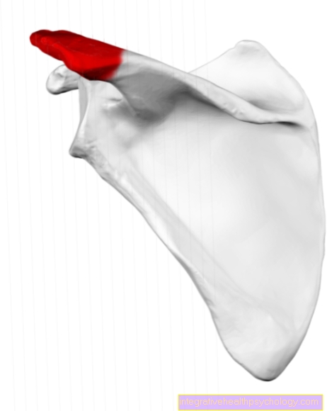
Simulation image of the left shoulder
Simulation of the irradiation
If the plan has been calculated and accepted after checking the quality requirements, an irradiation is now run through, i.e. simulated.
To do this, the patient is laid down in the same way as he was during the computer tomography. The alignment is done with the help of the skin markings.
When calculating the radiation plan, images were reconstructed that show what X-ray images would have to look like if they were made from the angles from which the linear accelerator shines. With the help of a special X-ray device, the X-ray simulator, X-ray images are now made from these angles and these are compared with the calculated ones. If these match from all angles, the radiation therapy can be carried out in this way.