Lung cancer diagnosis
diagnosis

If bronchial carcinoma is suspected, an X-ray overview of the lungs usually provides initial information - and possibly a suspicious result.
Read more on the topic: Chest x-ray (chest x-ray)
Further examinations to confirm the diagnosis or to rule out lung cancer are in particular computed tomography and bronchoscopy (reflection of the airways) with the removal of tissue samples (biopsy).
The diagnosis of lung cancer is often complicated because the symptoms are initially very unspecific. Here you can find more information on the topic: How do you recognize lung cancer?
If a tumor is found, a series of further examinations (usually in the hospital) are necessary in order to determine the extent of the disease and to record any concomitant diseases.
The diagnosis then serves:
- the exact location of the tumor (usually by computed tomography and chest x-ray)
- the histological classification (see also under epidemiology, mostly by bronchoscopy)
- the exclusion of distant metastases (usually an ultrasound examination of the abdomen, a computer tomography of the head and a skeletal scintigraphy are used here
- the assessment of operability (for this purpose, parameters are primarily collected that allow conclusions to be drawn about the function of the lungs)
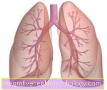
Bronchoscopy
The bronchoscopy is used to examine the airways, i.e. the windpipe and the large branches (bronchi). The term bronchoscopy is made up of the two Greek parts of the word "bronchus" (windpipe) and "skopein" (to look).
With a special endoscope (bronchoscope), a device that consists of a flexible tube and a camera at the tip, the doctor can view the airways from the inside and thus e.g. Detect endobronchial (broken into the bronchi) tumor growth. The bronchoscope is also equipped with a working channel for surgical instruments through which tissue samples (biopsies) can be taken. This allows cells to be obtained directly from the tumor tissue, which can be used to determine the type of tumor present.
To detect tumors that are inaccessible to the endoscope, the doctor can also perform a bronchial lavage. The bronchi are flushed with saline solution. After rinsing, the solution is then examined in a laboratory for tumor cells, fungi or inflammatory lung diseases.
Endosonography
In endosonography, a specially shaped ultrasound head is inserted over the esophagus. This makes it possible to focus on the areas around the airways Lymph nodes view, assess their size and, if necessary, perform a puncture and thus remove cells directly from suspicious lymph nodes in order to confirm or rule out an infection.
Check lung function
The examination of the lung function (see asthma) should show to what extent the lungs are still efficient. If part of the lungs or even an entire lung has to be removed, lung function will deteriorate. So if there is a severe restriction in advance, the operation cannot be performed.
Positron emission tomography PET
PET is a relatively new imaging technique that can be used to visualize cells with increased metabolic activity. The process is characterized by a high level of sensitivity, e.g. Currently a tumor from a size of 1-2 cm is detectable.
Read more on the topic: Positron emission tomography
histology
Histology (tissue examination) describes the cell composition of the tumor, on which the prognosis and treatment options depend. In bronchial carcinoma, a distinction is made between small-cell and non-small-cell tumors.
The classification of lung cancer is possible with the help of histology. The most common types of lung cancer are adenocarcinoma and squamous cell carcinoma. We recommend our main page at this point: Squamous cell carcinoma of the lungs