Broken finger
introduction
Both Fingers anatomically, these are structures in our body that are very easily injured. Broken fingers are one of the most common traumatic events in the emergency room.
To understand the broken finger it helps to understand the basic anatomy of the hand to understand. The hand is divided into three areas: wrist, Palm, and finger.
The fingers are most commonly affected Hand injuries. They consist of each three bone parts: Phalanx proximalis, media and distalis, with only the thumb has only two phalanges.
All these bone can be affected as part of a broken finger. The structures are connected via Tapes and one multilayered muscles.
Despite these protective structures, finger bones are repeatedly broken in Germany.

In the event of broken fingers proximal (the bone behind the metatarsophalangeal joint), medial (middle finger bone) and distal (the bone under the nail) Finger fractures distinguished.
The distal fracture is a more common one Sports injuries, it accounts for almost half of all hand fractures. Usually the middle finger is affected, the causes are often traumatic movements such as twisting or overstretching as well Bruises attributable to. Fractures in the phalanx are permanent forces of many Hand muscles exposed and for this reason very susceptible to twisting or shortening and can stick out in non-physiological angles.
Causes of a Broken Finger
The causes for breaking a finger bone extremely variable. Many everyday movements demand the activity of our fingers.
Violations of the Finger bones usually arise in the context of sporting activities, work or traffic accidents or others traumatic events.
The reason for breaking a finger bone can be one excessive stretching, twisting, bruising, or a direct blow be on the finger.

I would be happy to advise you!
Who am I?
My name is I am a specialist in orthopedics and the founder of .
Various television programs and print media report regularly about my work. On HR television you can see me every 6 weeks live on "Hallo Hessen".
But now enough is indicated ;-)
In order to be able to treat successfully in orthopedics, a thorough examination, diagnosis and a medical history are required.
In our very economic world in particular, there is too little time to thoroughly grasp the complex diseases of orthopedics and thus initiate targeted treatment.
I don't want to join the ranks of "quick knife pullers".
The aim of any treatment is treatment without surgery.
Which therapy achieves the best results in the long term can only be determined after looking at all of the information (Examination, X-ray, ultrasound, MRI, etc.) be assessed.
You can find me at:
- - orthopedics
14
Directly to the online appointment arrangement
Unfortunately, appointments can only be made with private health insurers. I ask for understanding!
Further information about myself can be found at -
diagnosis
As a rule, if a fracture of a finger bone is suspected, a X-ray image made to confirm the diagnosis if necessary.
On the X-ray, the Type of break can be recognized and the therapy tailored to the individual case.
Because it various forms of fractures If there are any other therapy options, an exact diagnosis by the attending physician is absolutely necessary in the event of a broken finger.
Since the bones of the finger are relatively small, it may be that several X-rays have to be taken in order to identify exactly whether and which fracture is present. To make a comparison, it can also be that the unaffected hand needs to be x-rayed.
Carrying out a Computed tomography (CT) is usually only carried out for very complex bone fractures. With the help of computed tomography it is possible to assess fractures that cannot be reliably diagnosed in an X-ray.
A disadvantage of this technology that should not be underestimated is that it is clear higher radiation exposure for the examined.
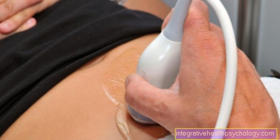
With the help of Magnetic resonance imaging (MRI; magnetic resonance imaging) diagnose broken bones that are not visible on a regular x-ray. This technique is also useful for assessing neighboring ones Blood vessels and cartilage structureswhich is also not possible in an X-ray image.
If the diagnosis of a broken finger is not established through the diagnosis, it can also be a bruise of the finger.
You can find more information on the topic here: Bruise of the finger
Duration of a broken finger
The duration of treatment for a broken finger can vary due to the various manifestations of this type of injury. For most cases, however, some guideline values can be formulated.
The affected finger should first (possibly after surgical treatment) with the help of a rail or one Plaster cast over a period of immobilized for about 3-4 weeks to give the two parts of the bone enough time and rest to grow back together. On it subsequently should be about again as long a periodin which the finger uses a Tape bandages is largely immobilized. In a sense, this represents a compromise between stability and mobility to be regained, since it is already back slight movements of the finger allowed.
Since the fingers are parts of the body that are extremely frequently used in everyday life, immobilization over such a long period is often particularly difficult. Many patients do not succeed in constantly recalling patience and giving the finger the time it needs to heal. In such cases, impaired healing of the fracture can occur, which can result in protracted complaints. Likewise, inadequate coalescence of the fracture point in the later course can under certain circumstances promote osteoarthritis in the finger. From these aspects it becomes clear why a sufficiently long rest period is so important for the affected finger.
Mobility after a broken finger
Due to the long immobilization of the finger, almost all patients with a broken finger develop a more or less strong one Restriction of mobility of the affected finger. To counteract this, you should already after removal of the splint or plaster cast with more targeted physical therapy to be started.
The therapist tries to do this Fingers carefully to mobilize. This can certainly lead to pain in the finger, but this can and must be accepted to a certain extent. The mobilization by the physiotherapist can be done well combine with the application of a tape dressing, as the therapist has special expertise and experience here too, which can have a positive effect on the further course of the finger's mobility.
The patient should be given a detailed explanation of the extent to which he can move the finger in everyday use of the affected hand and which Exercises to improve mobility of the finger too at home itself can perform. It is easy to see that restricting the mobilizing exercises to the physiotherapy units, which usually only take place twice a week, cannot provide a sufficient level of training and that these units should therefore be supplemented by independent units in the home environment.
Frequency distribution
Overall is a Fracture of the finger bones a very common reason for presenting to an emergency room. The most common is Terminal bone, so the Phalanx distalis affected.
A Canadian study gives 0.29%, i.e. 29 per 10,000 people over the age of 20, and 0.61%, i.e. 61 per 10,000 people under the age of 20, of the incidence, i.e. how many new finger fractures occur each year, who seek medical treatment every year for a broken finger.
The same study also shows that the male sex with 64% has an increased risk of broken fingers.
This is especially true in the age range between 20 and 60 years due to increased risk factors in individual behavior.
From the age of 65, women lead to the development of finger fractures, probably due to a lower stability of the bone. According to the study, young women aged 10-14 have an increased incidence of finger fractures, which is explained by a fragile bone structure due to the growth phase.
Symptoms
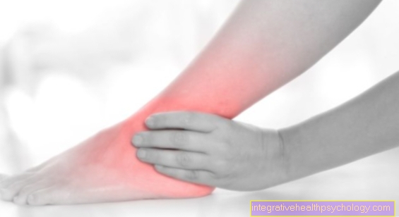
The symptom a broken finger is the entering of pain immediately after the injurious event.
In some cases the break can be seen directly from the outside if the Deformed fingers is.
Depending on the fracture, the affected person may still be able to move the finger, albeit with severe pain. Depending on the location and type of bone fracture and the associated stability, some will Broken bones described as painful than others. After a while, mostly within 10 minutes to step Swelling on the affected finger and the mobility of the finger decreases.
Depending on the extent, swelling can also affect surrounding fingers. In most cases, drowsiness of the finger also occurs, which is due to the compression of the surrounding area annoy can be explained by the swelling.
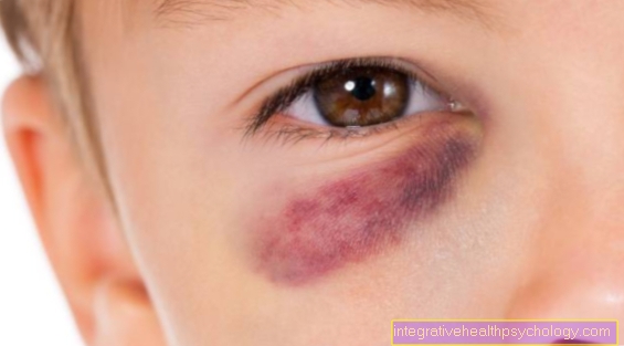
Depending on the person concerned bone it can also, if the Phalanx distal, so the bone under the Fingernail, is affected, come to a red fingernail because there is a Bleeding (hematoma) can form.
In very extreme cases it can also be that the bone penetrates the structures to the outside and is now visible from the outside.
Pain and swelling
Fractures are generally painful, and so are broken fingers. A special pain stimulus is given when the Periosteum, the periosteum, tears. It comes to the appropriate place Hemorrhage and for the release of Inflammation mediators. These are certain messenger substances that are responsible for the typical characteristics of inflammation. These include the increased Pressure and temperature sensitivity, Pain and swelling. This comes about because the Vessels at the injured area become permeable and water can escape into the surrounding tissue. On the distal phalanx there is a fibrous structure that bone and skin connects with each other or a network of very small compartments (separations) forms.
In the event of a fracture in the distal phalanx, the fingers can be located in these compartments blood and collect fluid, which can lead to further swelling and severe pain. Since the Nail bed is close to the bone, there can also be corresponding injuries, such as for example Nail tear or too painful Bruising (Hematomas) (please refer: Bruise under the nail). In order to prevent swelling or to recede initially, all fractures should first be covered with ice chilled and be stored high so that less blood or fluid escapes (store against gravity)
Therapy broken finger

The Goal of every therapy after a finger fracture consists in correcting the condition before the fracture as anatomically accurate as possible and the Mobility of the finger to ensure, so the Function of the finger restore.
This can be done with a variety of different therapies, each iindividually tailored to the injury will take place.
In any case, it is important to have one Broken finger to treat, otherwise Hand dysfunction can arise in the long term.
For this reason, it is recommended that you seek medical attention as soon as possible after a fracture of the finger bone.
In most cases, a operative therapy recommended to have a surgeon die Bone ends with screws or a wire reconnects.
It must be ensured that the soft structuresthat surround the bone are spared as possible during the operation.
In most cases, a sufficient stability after 3 to 4 weeks to be guaranteed.
In the case of fractures of the distal phalanx, i.e. the Phalanx distalis, one is enough in most cases rail which immobilizes the finger and usually allows healing after a few weeks.
Bleedingthat often occur with this type of broken finger should be removed to remove the Fingernail to relieve. In some cases, however, this fracture may require surgery to repair free pieces of bone to fix.
Exercise should be started as soon as possible after the surgical treatment in order to guarantee the absolute functionality of the finger. The individual point in time for the start of a physiotherapy therapy is decided by the attending physician after assessing the finger after the operation.
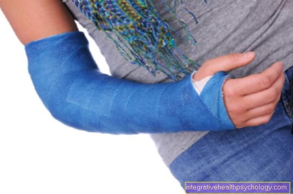
Splint / plaster of paris for a broken finger
Non-displaced (displaced) fractures are immobilized with a splint for at least three to four weeks.
In the case of fractures of the distal phalanx, the splint should extend beyond the fingertip to protect the finger from further injuries. There are splints that hold the distal interphalangeal joint (the finger joint closest to the nail) so that the other joints remain flexible. This prevents stiffening of the other phalanxes.
Most fractures heal well with conservative therapy, typically three to four weeks. More complicated shaft fractures often require prolonged immobilization. Even after splint / plaster therapy, the injured finger should be fixed on the neighboring finger for three to four weeks with tape or something similar and thus stabilized.
Read more about the topic here: Tap your fingers
However, under certain conditions, surgery is recommended instead of splint therapy. This is the case when tendons, nerves or vessels are damaged, the joint is directly affected or there are serious nail / bed injuries. A hand surgeon should also be consulted in the case of twisted, significantly angled, shortened or otherwise deformed fractures.
Taping a broken finger
Taping a broken finger only comes in extremely rare cases (especially with an uncomplicated fracture of the little finger) as the primary measure immediately after the injury in question. This is due to the fact that the stability guaranteed by the taping is simply too low to seem sufficient for a fresh finger fracture. Because of this, will first (or after a possibly necessary operation) with a rail or one plaster cast worked. Only when this over 3-4 weeks have made it possible for the fracture to heal optimally Tape replace them.
To do this, the affected finger is attached to an adjacent finger with the help of special medical tape. The patient should obtain the correct technique for this from a specialist, such as a Physiotherapists, explain and show. Otherwise, improper taping can force the finger into an incorrect position, or the tape does not create a sufficient degree of stability and thus endangers the long-term preservation of the previously achieved treatment success.
Surgery for a broken finger
The decision on the surgical treatment of a broken finger is made taking various factors into account. First and foremost, this includes the question of whether the broken finger is a simple one or one complex break with several fragments acts. In the latter case, the decision to have surgery is made much more frequently. Another important consideration relates to determining whether the individual Pieces of bone on either side of the fault line shifted against each other are or are still in their anatomically correct position. A displaced fracture should always be treated with surgery, unless the displacement can be corrected by repositioning maneuvers. This is followed by a similar therapy in the form of immobilization as with an unshifted broken finger.
The is a special case with regard to the therapy of a broken finger open finger fracture This is, by definition, if the Skin at the point where the finger was broken is no longer intact and the bone is thus more or less exposed. As an open break as possible Entry gate for pathogens can serve, it should be tackled operationally immediately. First the bone fracture is treated and then the adjacent soft tissues are reconstructed as much as possible. Finally, the skin defect is closed to avoid infection.
The surgical treatment of a broken finger is one comparatively simple intervention. The surgeon connects while the two Bone parts with a screw or wire. In addition, possible accompanying injuries, such as Tears in the finger tendons or bleeding under the fingernail, should be corrected. These are often the decisive aspect for performing an operation in the first place, since they rarely heal on their own without complications than the broken finger itself.
prophylaxis
A Broken finger usually happens due to an accident. Risk factors are these Performing contact sports how Hockey, soccer or Handball, but also certain professional groups fall under the risk profile for acquiring a broken finger.
People in these risk groups should therefore pay particular attention to their fingers and should not underestimate the risk of a finger fracture.
forecast
The forecast after a broken finger depends on many different factors. The timely diagnosis and the early start of therapy play a major role in assessing the prognosis.
After a long period of time without therapy, a broken finger can be expected to cause damage over the long term. This is also the case when a Immobilization after an operation took too long. This is why you should use it as soon as possible after an operation function-maintaining exercises to be started.
cure
Patient with uncomplicated stable fractures can already after one to three weeks start moving your finger again to avoid stiffening the finger.
After approx. four weeks should the old one Mobility and freedom from pain be guaranteed, but caution is required when exercising.
At more complicated, dislocated fractures it can Months take time for the bones to grow back together, usually 6 to 12 weeks. The duration of therapy is determined by visible healing on the X-ray image as well as regaining full and pain-free mobility of the finger.