Blood vessel
Synonyms: Vas sanguineum, vein
definition
A blood vessel is a hollow organ with a specific cell structure that is typically made up of several wall layers.
Blood vessels form a coherent system in the body for the transport of blood, the bloodstream.
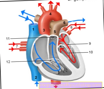
They are responsible for the entire transport of oxygen and nutrients in the body. They form a complex system of strong arteries with thick wall layers up to small, fine capillaries. In the following, the subdivision and tasks of blood vessels will be discussed in more detail. It is important to remember, however, that arteries always flow away from the heart and veins towards the heart. The often known division into vessels with oxygen-rich or oxygen-poor blood is in fact incorrect with regard to the large and small body circulation.
The total length of all blood vessels in the human body can be up to 150,000 kilometers. It can also be said that blood flows in almost every area of our body. Exceptions are the cornea in the eye (Cornea), the enamel, hair and nails.

Classification

Blood vessels can be subdivided again depending on their size and the blood to be transported.
The largest vessels are the arteries. You will be over Arterioles smaller and smaller down to the capillaries.
After all, capillaries have the smallest diameter and only have a very thin wall structure.
They are therefore particularly well suited for gas exchange in the lungs.
The venules, which are responsible for the transport of oxygen-poor blood, then connect to the capillaries.
Vessels of this type with a larger lumen are called veins.
The main artery, also known as the aorta, as the largest artery in the body, and the upper and lower vena cava (Vena cava superior and inferior), which carries the collected blood back to the heart.
Furthermore, a distinction is made between a muscular and an elastic type among the arteries.
Muscular-type arteries form the largest group.
In contrast, the arteries near the heart, such as the aorta and the large pulmonary vein, are of the elastic type.
Read more on the topic: Artery types
function
The blood vessels and that heart collectively as pumping organs, they form the body's blood circulation.
All organs, such as the head, legs and arms, pass through the bloodstream With Blood and the dissolved in it Provides nutrients and oxygen.
Be at the same time Degradation products, Metabolic waste products and carbon dioxide transported away and about the heart back to the lungs led to re-oxygenate the blood there.
The place for gas and mass transfer are the capillaries.
You are for it particularly well suitedas they have a low layer thickness and because of her thin diameter have a slow flow velocity.
Through the slow blood flow enough time remains in the capillaries to allow diffusion oxygen from the inhaled air record and at the same time Release carbon dioxide.
Air vessels
As Air vessels the great arteries are called aorta and their branches.
They typically contain a high proportion of elastic fibers and are therefore part of the elastic type.
Through the Air vessel function becomes the pulsating flow flowing through the irregular pump performance of the heart, in the more distant arteries more and more into one continuous flow transformed.
This is done in that during the systole only about half of the blood flows directly into the arteries. The other half is initially stored in the enormously elastic aorta.
The Wall of the aorta possessed by the numerous occurring elastic fibers very good restoring forces, which then push the stored blood into the arteries during diastole.
This compensates for pressure and flow peaks.
Resistance vessels
Small arteries and the arterioles are called resistance vessels.
They are used to reduce blood pressure before it enters the capillaries.
In their totality they make up 50% of the total resistance.
This effect is based on the sharp decrease in the individual diameter of the vessels.
The total resistance is very strongly influenced by this and has a great influence on the total peripheral (distant from the heart) resistance.
An important vessel or vascular section that ensures a continuous flow of blood is the root of the blood. It is a few centimeters long and plays an important role in the air chamber function.
If you are more interested in this topic, read it below: Aortic Root - Anatomy, Function & Diseases
Capacity vessels
As Capacity vessels one describes the parts of the venous system.
The veins have a very good one Compliance. Compliance describes the property of a vessel to take up a certain volume due to elastic fibers, despite a slight increase in pressure.
As a result, the capacity vessels are able to approximately 80% of the total blood volume save. If necessary can this volume mobilized by the tone vascular smooth muscle is increased.
Sphincter vessels
Vessels of this type have one ring-shaped locking mechanism. Through this it is possible to use the Blood flow the downstream Arteries to regulate.
For example, the arterioles control the flow of blood into the Capillary system.
Capillary system
Ultimately, it remains to be said that Capillaries for the Mass transfer are responsible. While fat soluble substances free themselves through the wall have to move water-soluble substances "diffuse" through the wall or use other transport systems. Since the capillaries are of enormously important physiological importance, it is useful to know their subdivision:
Continuous capillaries
fenestrated capillaries
Sinusoid capillaries
Continuous capillaries:
With continuous capillaries, the cells usually form one completely closed wall.
Fenestrated capillaries:
This type of capillary has Pores in its inner layer on that for the Exchange of low molecular weight substances are important. They come mostly in Intestinal tract before, since the absorption capacity is relatively high here.
Sinusoid capillaries:
Sinusoids or also discontinuous Own capillaries a significantly enlarged vessel diameter than the other two types of capillaries. They also own a lot large pores. Even large molecules such as proteins can be absorbed through this wall.
construction

Most vessels have a characteristic three-layer wall structure. This can vary depending on the type of vessel and the conditions.
In general, the higher the mean pressure, the thicker and more muscular the middle layer of the vessel.
Innermost layer (Intima)
The innermost layer is made up of a single-layer cell structure, also known as Endothelium referred to as.
These cells are aligned lengthways so that they can ensure a smooth flow of blood through the vessels.
The Endothelium sits on a basal layer, the Basal lamina. She anchors that Endothelium with the muscle cell-rich layer below.
Under the Endothelium lies the so-called subendothelial layerthat mostly made up Extracellular matrix exists, i.e. connective tissue, and hardly contains any cells.
The veins have a special feature in this layer. The venous valves, which promote the return flow of blood to the heart, form a duplication of the intima and close like a valve when the flow is reversed.
Middle Layer (Media)
The Media is the thickest layer of the vessel wall and is covered by the Intima through the so-called Membrana elastica interna separated, a fiber-rich thin layer that promotes mobility. It mainly contains smooth muscle cells and Extracellular matrix with elastic and collagen fibers.
The circular muscle cells are used to regulate the vessel size.
To the Media In larger vessels, a so-called often closes External elastic membrane on.
Outer layer (Adventitia)
The outer layer is a connective tissue layer that embeds the vessel in the surrounding tissue. It contains, among other things Fibroblasts, elastic fibers and collagen fibers. In addition, there are the smallest vessels to supply the arteries (Vasa vasorum) and lymph vessels.


