Representation of the paranasal sinuses using MRI
introduction
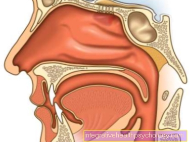
The paranasal sinuses are cavities within the facial bones of the skull that are filled with air, arranged around the nasopharynx and lined with nasal mucosa.

They are divided into the so-called
- Maxillary sinuses
- Frontal sinuses
- Sphenoid sinus
and - Ethmoid cells,
communicating with each other and with the nasal passages of the nasal cavity.
They serve primarily as a Resonance spaces when speaking as well as for the Humidification, cleaning and heating of the air.
MRI or CT imaging of the sinuses?
The Magnetic resonance imaging (MRI) as an imaging process that does not work with harmful X-rays but with magnetic fields is particularly suitable for Soft tissue imaging and thus also for imaging pathological processes (e.g. Sinus infection, Tumor formation) the Sinusesin which there are i.a. the mucous membrane is involved.
The Computed tomography (CT), Another method for displaying the paranasal sinuses can also be used to assess the mucous membranes, but is also particularly suitable for bony imaging (e.g. to clarify the anatomical conditions in the paranasal sinus system). However, in contrast to the MRI, it works with X-rays.
Both procedures can be used equally, depending on where the focus is on the question and which indication is behind the examination.
You can find a lot more information under our topic: MRI or CT -What is the difference?
Indications
The MRI (and also the CT) is used in the imaging of the routine general diagnosis of the paranasal sinuses in the facial skull, especially Inflammatory processes and Masses of the nasal mucosa, Facial skull bone changes and the anatomical features of the sinus system can be assessed.
The most common indications for an MRI of the paranasal sinuses is sinusitis (med. Sinusitis). Especially in the case of chronic sinusitis, the MRI can provide information about the cause of the chronification, e.g. by evidence of outflow prevention etc.
In addition, the MRI display is also used for imaging before planned invasive surgical interventions on the sinuses, such as Punctures or endoscopies.
In general, these are common indications for a Sinus MRI of the Exclusion of inflammatory or space-consuming processes such as Follow-up checks in this regard, Representations of congenital anatomical variants and malformations, overview images before surgical interventions and the exclusion of fractures after trauma.
In particular, different Differential diagnoses represent an indication for an MRI:
- therefore belong to this Inflammation, such as acute or chronic sinusitis (sinus infection)
- Accumulation of mucus or pus in the cavities
- Fractures in the midface or frontobasal (skull base fracture, frontal bone fracture) after trauma
- benign tumors (e.g. osteomas, polyps, angiofibromas, retention cysts)
- malicious Tumors (e.g. carcinomas, sarcomas, metastases)
- congenital malformations, such as a narrowing or occlusion of the posterior nostril (Choanal stenosis, atresia), one Cleft lip and palate or that Kartagener's syndrome
MRI for a sinus infection
As part of the diagnosis of a suspected Sinus infection closes that MRI as further diagnostic imaging, usually a physical examination, a Taking a smear from the nasal secretions and one Rhinoscopy (Nasoscopy), but mostly only if complications arise, a surgical intervention for therapy is planned or a chronic course of sinusitis is present.
Simple, acute, uncomplicated courses usually do not require any further diagnosis by means of an MRI.
Procedure for the head MRI examination
Special preparation for the MRI examination is usually not necessary, it should only be ensured that There is no more food or fluid intake approx. 4 hours before the start of the examination.
Loose clothing is recommended on the day of the examination without metal parts (e.g. buttons, zippers, underwired bra, etc.), these can usually be left on during the examination.
The patient is also required to remove all metallic objects from the body (e.g. jewelry, watches, teeth, piercings, hair clips, etc.).
Then the patient lies supine on the examination table, which is then pushed into the MRI machine head first. If a patient suffers from claustrophobia, a sedative can be administered beforehand.
For this and also for a possibly necessary administration of a Contrast agent before or during the examination, a Indwelling cannula be placed in the elbow-arm vein.
Sound during the examination loud knocking noisesthat caused the MRI. If this is perceived as uncomfortable or annoying, headphones can be given to the patient, via which music can be played and the staff can also contact the patient.
In addition, each patient receives a Emergency bell in the hand, which can be pressed at any time during the examination, should problems arise.
The MRI examination lasts in total about 20 minutes, in which sectional images with the help of magnetic fields of the head and sinuses are made while the patient is stay as calm as possible should.
MRI examination with contrast agent
The administration of a contrast agent enables a more precise and better representation and image quality, so that it can be used before or during the examination.
The contrast medium, which is given into the bloodstream via the vein, is particularly concentrated where the blood flow is increased, e.g. also for sinus infections or tumors.
The contrast media for an MRI examination do not contain iodine and are usually well tolerated without any side effects.
Please also read our topic: MRI with contrast agent


