The elbow joint
Synonyms
Medical: Articulatio cubiti
definition
The elbow joint (Articulatio cubiti) connects the upper arm with the forearm. It consists of three partial joints, which are formed by three bones (upper arm, ulna and radius):
- Humeroulnar joint (Articulatio humeroulnaris):
formed by the upper arm (humerus) and ulna - Humeroradial joint (Articulatio humeroradioalis):
formed by the upper arm and spoke (radius) - Proximal Radioulnar Joint (Articulatio radioulnaris proximalis):
formed by the proximal ends of the ulna and radius (close to the body)
These partial joints are combined with a common joint capsule to form the elbow joint.

Figure elbow joint

- Humerus head -
Capitulum humeri - Outer femoral gnar -
Epycondilus lateralis - Inner femoral gnar -
Epycondilus medialis - Upper arm roll - Trochlea humeri
- Upper arm shaft -
Corpus humeri - Spoke head - Caput radii
- Spoke neck - Collum radii
- Roughness of the spoke -
Radial tuberosity - Roughness of cubit -
Ulna tuberosity - Spoke shaft -
Corpus radii - Ellschaft -
Corpus ulnae - Articular cartilage
- Joint capsule -
Articular capsule - Celiac nerve -
Ulnar nerve - Arm extensor -
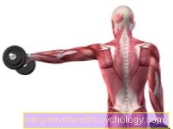
Triceps brachii muscle - Upper arm muscle -
Biceps brachii muscle
You can find an overview of all Dr-Gumpert images at: medical illustrations
function
The elbow joint can be in two degrees of freedom be moved.
On the one hand, the forearm can be opened when the upper arm is not moving bend and stretch (Flexion / extension).
On the other hand, the elbow joint is functionally connected to the proximal radioulnar joint Turning movements involved in the hand (Pronation/Supination).
The main movements in the elbow joint are controlled by the Upper arm muscles executed.
The flexors (Flexors) are located on the front of the upper arm. These include:
- the Biceps brachii muscle (Biceps)
- and the Brachioradialis muscle (Upper arm spoke muscle).
The extensors in the elbow joint are on the back of the humerus. Also includes:
- the Triceps brachii muscle (Triceps).
Individual forearm muscles are then also involved in pronation and supination.
Appointment with a tennis elbow specialist?

I would be happy to advise you!
Who am I?
My name is I am a specialist in orthopedics and the founder of .
Various television programs and print media report regularly about my work. On HR television you can see me live every 6 weeks on "Hallo Hessen".
As a former performance-oriented tennis player, I specialized early on in the conservative treatment of chronic tennis elbow.
In the past few years I have successfully treated several thousand tennis arms.
You can find me in:
- - your orthopedic surgeon
14
Directly to the online appointment arrangement
Unfortunately, it is currently only possible to make an appointment with private health insurers. I hope for your understanding!
You can find more information about me at .
Humeroulnar joint
in the Humeroulnar joint (Articulatio humeroulnaris) form the "role" of the upper arm (Trochlea humeri) with a corresponding indentation on the ulna (Trochlear notch) a joint.
In close spatial connection to Trochlear notch, it says Olecranon, a protruding bone of the ulna that can be palpated as an "elbow".
The humeroulnar joint enables this Flexion and Extension (Flexion and extension) and is therefore a so-called Hinge joint.
Humeroradial joint
The Humeroradial joint (Articulatio humeroradialis) is through the connection of the humerus head (Capitulum humeri) and the corresponding specialization (Fovea articularis radii) on the head of the spoke (radius head, Caput radii) educated.
This joint also has the two degrees of freedom the Flexion / extension to Flexion and extension of the forearm and the Supination / pronation to rotation of the hand.
Strictly speaking, this is a Ball joint. Ball joints always have three degrees of freedom (In addition to the degrees of freedom of the hinge joint, the abduction and adduction).
Since the humeroradial joint, however, by very strong band connections is secured, this last degree of freedom is omitted, so that it is anatomically a ball joint, which, however, only two degrees of freedom owns.
Proximal radioulnar joint
in the proximal radioulnar joint (Articulatio radioulnaris proximalis) are the edge of the radius head (Circumferentia articularis radii) and the corresponding notch on the inside of the Ulna (Incisura radialis ulnae) articulated together.
They form a so-called Wheel jointwhich one Rotation around the longitudinal axis of the bones makes possible. So that is this joint essential to the turning and turning movements of the hand involved.
Joint capsule and strap securing device
The common large joint capsule closes all three partial joints and thus functionally combines them to form the elbow joint.
Attached is the joint capsule all three involved bone, i.e. on the upper arm, the spoke and the ulna.
In the area between the ring ligament (explanation follows) and the neck of the radial head, the joint capsule forms a bulge, the so-called protrusion Sacciform recess. This excess of capsular tissue serves as the Reserve fold and is used when the forearm is fully rotated in one direction.
The humeroulnar and humeroradial joints have strong band connections (Collateral ligaments) that lie laterally on the joint capsule.
These tapes (Ligamentum collateral ulnare and Radial collateral ligament) run in strong, fan-shaped stripsso that they support the joint laterally in every position:
- The Lig. Collateral ulnare pulls from the protruding bone above the central upper ambone (Epicondyle medialis humeri) to the joint attachment of the upper arm to the ulna (Trochlear notch)
- The Collateral radial ligament arises from the protruding bone above the lateral humerus (Lateral epicondyle of the humerus) and then pulls into the ring band.
The Ring band (Annular radial ligament) originates from the ulna, moves around the radius head and starts again from the ulna. This is how it secures the proximal radioulnar joint.
Bursa
Bursa are liquid-filled, capsule-like delimited cavities, the outside the joint space lie and cushion heavy mechanical loads.
Bursae are either congenital or acquired (reactive bursae). Depending on the mechanical stress, bursae of different sizes develop in different places in each person.
Through this high individual variability you can no details to make the bursae of the elbow joint.
The largest bursa in the elbow joint is called Bursa subcutanea olecrani. It lies between the top of the ulna and the skin.
In the event of high mechanical stress or an open wound, a Bursitis come.
Diseases
- In the Epicondylitis it is an inflammatory and painful irritation between the visual attachments of the muscles and the protruding bones to which they are attached. These are caused by overuse and are popularly called "tennis elbow" or "golfer's elbow" in the elbow area, depending on their location.
This disease can be treated with immobilization and the administration of painkillers and anti-inflammatory drugs. - The olecranon bursitis is also caused by severe mechanical irritation and overuse. It is an inflammation of the bursa in the subcutaneous fatty tissue of the elbow.
This inflammation is treated by immobilizing the joint, reducing mechanical stress, cooling and administering anti-inflammatory drugs. In chronic and severe cases, the bursa may even have to be surgically removed.
Read more on the subject: Tendinitis in the elbow.
- The Ulnar groove syndrome caused by pressure damage to the ulnar nerve in the area of the elbow. It runs through a palpable groove on the inside (sulcus ulnaris) and is only protected by a small amount of fat and skin tissue. Tingling, paresthesia, pain and even paralysis are among the symptoms. Initially, an attempt is made to provide relief through relief, padding or splinting, but sometimes surgical intervention is required and the nerve has to be freed from its too narrow tissue bed (neurolysis).
- In addition to joint inflammation (arthritis), various fractures (Fractions) and Dislocations (Dislocation of the joint), one form of subluxation occurs quite often in small children: in the Chassaignac palsy Tension on the child's elbow joint, which is still quite unstable, results in a partial dislocation of the radial head, in which the annular ligament (ligamentum annulare radii) is pinched between the radial head and the humeral capitulum. This leads to severe pain when moving, which is why the children adopt a typical gentle posture with flexion and inward rotation. The ring ligament can be brought back into the correct position very quickly by stretching it and turning it outwards while pulling it. No further immobilization or treatment is necessary.
- A tear in the ligamentous apparatus of the elbow joint can also occur. Such a ligament tear is often caused by jerky movements.
Read more on the topic: Torn ligament on the elbow