The conduction anesthesia at the dentist
Conduction anesthesia is a local form of anesthesia in which certain nerves or nerve branches are numbed during an operation. In the case of dentistry, this eliminates the pain in larger intraoral areas. Conduction anesthesia is possible in both the upper and lower jaw.

Reasons for block anesthesia
With conduction anesthesia, a larger area is often anesthetized. This can be particularly desirable for larger interventions. For example, it may be possible for the dentist to work on several teeth or gum areas in the course of a treatment. In order to ensure the patient is as free from pain as possible, conduction anesthesia is usually chosen.
It is possible to numb larger areas of the gums or palate and several teeth at the same time. Another crucial point for the choice of conduction anesthesia is the bone structure in the lower jaw. Due to the very compact (i.e. dense) bone structure in the lower jaw, so-called infiltration anesthesia, which is otherwise the method of choice, cannot achieve the desired depth of anesthesia. The local anesthetic cannot achieve the desired effect and the patient may have a painful, unpleasant treatment. In summary, the following examples can be given for the choice of central anesthesia:
- Major operations in the maxillary anterior region (infraorbital nerve),
- The removal of a mucosal graft from the palate (N. palatineus major),
- Treatments in the lower jaw (N. alveolaris inferior),
- Removal of wisdom teeth in the lower jaw
Also read: Gum transplant, Conduction anesthesia,
procedure
In the case of a conduction anesthesia in the upper jaw, the process hardly differs from that of an infiltration anesthesia. Please note the exact injection sites, which are not discussed in detail here.
It is worth mentioning that with anesthesia in the infraorbital foramen in the upper jaw there are also situations in which the syringe is placed extraorally (outside the mouth). This is especially the case if the patient is suffering from a so-called lock jaw due to certain injuries and is unable to open his mouth.
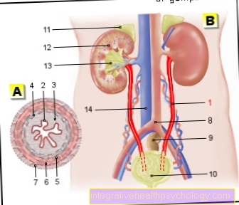
The exact structure of the upper jaw can be found here: Figure upper jaw
The sequence of anesthesia in the lower jaw will be discussed in more detail at this point.
The difficulty for the dentist is that the mandibular foramen is sometimes difficult to locate. The doctor has to deal with the individual anatomical circumstances in order to ensure adequate anesthesia.
- The doctor should guide the cannula about 1 cm above the rows of teeth, from the premolar region (the two teeth after the canine) on the opposite side towards the cheek.
- The puncture point is lateral to the so-called pterygomandibular plica, roughly in the middle between the upper and lower teeth.
- The dentist has to advance the cannula until it is in contact with the bone and should check whether he has hit a blood vessel before applying the anesthetic. If this is the case, a new puncture must be carried out so that hematoma formation from hitting the blood vessel can be avoided.
You might also be interested in the following articles: Local anesthesia at the dentist, local anesthesia
How painful is that?
As with all other forms of anesthesia, conduction anesthesia causes typical puncture pain. This can be a bit more uncomfortable with central anesthesia in the upper jaw, because the mucous membrane on the palate is particularly thin. This is why there is greater pain during anesthesia in this area, as the sensitive periosteum is irritated by the cannula.
It is possible to reduce the puncture pain with surface anesthesia. A spray is used here that is applied beforehand to numb the affected gums a little.
It can also be very painful if the dentist hits a nerve while piercing. The patients describe the feeling of a “lightning strike”. If this is the case, the dentist should definitely choose a new position for the application so that the nerve is not damaged. Furthermore, painful hematomas can occur if there is bleeding in the area of the puncture site.
What are the risks
With every form of anesthesia there are certain risks that the patient must be informed about in each case. Certain, very rare, risks are:
- Nerve damage possibly forever
- Cannula fracture
- Infections (injection abscess)
- Cardiac arrhythmias
- allergic reactions in the event of intolerance to the anesthetic
In addition, hematoma formation can occur if the local anesthetic is incorrectly delivered directly into a blood vessel. In most cases, however, these regress quickly.
A rare complication here is the jaw clamp, in which it is no longer possible to open the mouth due to the bleeding and the formation of hematomas. The jaw clamp also usually goes away without any problems after a few days.
In order to counteract the risks and to be able to guarantee a safe treatment, it is essential to collect a current anamnesis of the patient. Any intolerance or allergies, which are important for the choice of medication, may be noticeable here.
Please also read: Jaw clamp, abscess
How long does central anesthesia work?
The duration of anesthesia is usually between 1 and 5 hours. This depends on several factors
- On the one hand, the choice of anesthetic is important because, for example, the effect of Lidocaine only lasts 1-2 hours that of Bupivacaine however, up to 5 hours.
- In addition, the addition of adrenaline is crucial for the duration of the effect, since the anesthesia lasts longer when adrenaline is added. In many cases, however, adrenaline is indicated as an active ingredient and it should not be avoided for nothing.
- Also significant is that patients who regularly use drugs are more difficult to anesthetize. They usually need higher doses and the anesthetic wears off more quickly.
What does central anesthesia cost?
The conduction anesthesia is usually covered by the statutory health insurance, since the elimination of pain must be guaranteed during the treatment.
- According to the BEMA billing items, intraoral conduction anesthesia can be billed using item 41a and costs € 11.20. The extra-oral form (position 41b) costs 15 €.
- For privately insured patients, the intraoral conduction anesthesia can be billed according to item GOZ 0100 and costs € 9.05.
Differences in conduction anesthesia in the upper and lower jaw
As already described, it is usually sufficient to perform an infiltration anesthesia in the upper jaw, in which the teeth are anesthetized individually. Exceptions to this are major surgical interventions or mucosal grafts that have to be removed from the palate. A distinction is made between the following conduction anesthesia in the upper jaw:
- Tuberosity
- F. palatinum majus
- F. incisivum
- F. infraorbital
You can find out more about the anatomy of the upper jaw at: upper jaw
Due to the thicker bone structure in the lower jaw, conduction anesthesia is the method of choice for treating teeth. There are the following options:
- F. mandibulare (N. alveolaris inferior, N. lingualis)
- F. mental (N. mentalis)
- N. buccalis
You can find more about the anatomy of the lower jaw at: Lower jaw
It is important here that the inferior alveolar nerve is seldom anesthetized on its own, but that the lingual nerve is usually switched off due to the close positional relationship. This numbs the gums on the inside and the tongue as well.
With sufficient conduction anesthesia in the mandibular foramen, all teeth of the affected lower half of the jaw are numbed, as well as part of the gums and the tongue (lingual nerve). However, if you only want to numb the teeth of the lower jaw front, it is possible to perform anesthesia in the mental foramen. The nerve branch of the inferior alveolar nerve runs there and supplies this area. This makes it possible to only numb this area, which is often more comfortable for the patient, as the tongue and the back of the teeth can still react sensitively.
What can you do if central anesthesia does not work
There are various reasons why central anesthesia does not work. Mostly this is the case with anesthesia in the mandibular foramen in the lower jaw. Due to the difficult anatomical conditions and the individual nerve path of the patient, the anesthesia often fails.
If the dentist does not manage to find the necessary injection site, there is the option of performing what is known as intraligamentary anesthesia. The local anesthetic is injected directly between the tooth and the bone.
Many of the complications and risks can also be reduced by this form of anesthesia, as there are hardly any anesthesia failures and the risk of nerve or vascular damage is lower. Patients suffering from endocarditis (inflammation of the inner skin of the heart) should NOT receive intraligamentary anesthesia.
In complicated cases, the option of anesthesia at the dentist can also be considered.