Distal radius fracture
definition
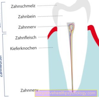
The distal radius fracture describes the fracture of the distal radius, i.e. the part of the spoke near the wrist.
With around 25% of all fractures, the distal radius fracture is the most common fracture in humans. It affects athletes as well as older patients who fall due to various causes. But postmenopausal changes in the hormonal balance can also promote a fracture. With a good 80%, the so-called Colles fracture is much more common than the Smith fracture.
In the Colles fracture, the fall takes place on the dorsally extended, upwardly extended hand. The fracture fragment is displaced dorsally and radially, i.e. towards the spoke.
The Smith fracture is, so to speak, the counterpart to the Colles fracture, and describes a fall on the downwardly angled, palmar-flexed hand. The fracture fragment is shifted palmar, i.e. towards the hand, and also radially (towards the spoke).

If, in addition to the radius fracture, there is also a dislocation of the ulna (ulna), one speaks of a Galeazzi fracture. Traumatologically, the cause here is a fall on the forearm turned outwards.
In addition to the two above-mentioned, very common forms of distal radius fractures, there are other less common fractures, which - named after their first description - have different names: In the case of a chauffeur fracture, the styloid process breaks off at the distal radius. Of the Radial styloid process is also called the stylus extension in German and describes a small extension near the wrist that laterally encloses the finger bones. In the Barton fracture, the upper part of the radial joint surface is also affected, so that - as in the case of the chauffeur fracture - one speaks of an intra-articular fracture, i.e. a fracture that includes the joint cavity. The anatomical, traumatological counterpart is the reversed Barton fracture, in which the lower part of the distal, radial joint surface is fractured.
Both Barton fractures involve the joint cavity or the joint and are therefore referred to as intra-articular.
causes
By far the most common cause of a distal radius fracture is falling on an outstretched arm. The arm is instinctively stretched out to catch the fall and, if necessary, to prevent worse. The resulting fraction is called a Extension fracture (also called Colles' fracture). A fracture can also result from falling on the bent hand - one then speaks of one Flexion fracture (Smith fracture). Distal radius fractures are particularly common in older patients, as their bone density is often affected by osteoporosis and is therefore more prone to fractures. With them, even minor trauma is sufficient to lead to a fracture that would not have led to a fracture in healthy patients. The second most frequent patient group, alongside older patients, are younger patients between the ages of five and eighteen. Sports accidents usually lead to a distal radius fracture. Traffic accidents can also lead to a forearm fracture.
Appointment with ?

I would be happy to advise you!
Who am I?
My name is I am a specialist in orthopedics and the founder of .
Various television programs and print media report regularly about my work. On HR television you can see me every 6 weeks live on "Hallo Hessen".
But now enough is indicated ;-)
In order to be able to treat successfully in orthopedics, a thorough examination, diagnosis and a medical history are required.
In our very economic world in particular, there is too little time to thoroughly grasp the complex diseases of orthopedics and thus initiate targeted treatment.
I don't want to join the ranks of "quick knife pullers".
The aim of any treatment is treatment without surgery.
Which therapy achieves the best results in the long term can only be determined after looking at all of the information (Examination, X-ray, ultrasound, MRI, etc.) be assessed.
You will find me:
- - orthopedic surgeons
14
You can make an appointment here.
Unfortunately, it is currently only possible to make an appointment with private health insurers. I hope for your understanding!
For more information about myself, see - Orthopedists.
Diagnosis
The diagnosis usually consists of a combination of a discussion with the patient in which the patient describes his symptoms and the accident, examination of the arm and a final X-ray examination of the arm. A distal radius fracture can only be definitively concluded through the X-ray examination - the patient discussion and examination are not sufficient for this. During the examination, which is usually only possible to a limited extent due to the patient's pain, the doctor pays attention to a misalignment of the arm, restricted movements, as well as sensory and circulatory disorders in the hand. In exceptional cases, if the doctor has the suspicion that surrounding ligaments or other structures may be injured, a magnetic resonance tomography examination (MRI) carried out. Computed tomography (CT) is rarely performed if multiple fractures are suspected.
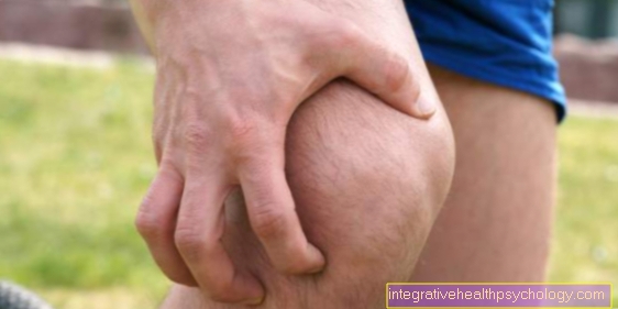
Pain
As in Broken bones common, pain also occurs with the distal radius fracture.
This is because when a bone breaks the fine Periosteum - the Periosteum - is pierced by the underlying bone tissue. The periosteum is, however, very strongly penetrated by small nerve fibers, which immediately send pain impulses to the brain send. The background is evolutionary in nature: a fracture had to be spared even in earlier times, and under no circumstances should it be further burdened, otherwise it would also Blood vessels or nerve tracts could be affected.
Only after weeks, when the fracture has healed, does the pain decrease, since an injury to the surrounding structures is now unlikely. In today's medicine, pain killers can of course be given to relieve pain so that the patient is symptom-free. However, it is then one "Deceptive peace", since the basic problem is of course not eliminated. Pain therapy only makes sense with simultaneous immobilization and surgical or conservative treatment of the fracture.
Because pain - as annoying as it may be - has you sense, as they signal the body to spare the affected part of the body. Preclinically freely available painkillers (medical: Analgesics) are pain relievers the NSAIDs-Group, such as Ibuprofen and Paracetamol.
In an acute case, an emergency doctor can also respond to low to high potency Opioids To fall back on. These are then administered intravenously and switch off pain very quickly. Painkillers are also usually prescribed for follow-up treatment. Aspirin® Like ibuprofen, it belongs to the NSAID class, but it also liquefies the blood, which is a nightmare for every surgeon. Vascular injuries are very difficult to breastfeed intraoperatively. Aspirin (generally acetylsalicylic acid) should therefore be avoided in the preclinical setting.
More symptoms
In addition to the pain to be expected, a distal radius fracture is usually accompanied by other symptoms. It is typical that the hand can no longer be properly loaded and the muscle strength is significantly reduced. The hand is usually held in a relieving position due to the pain. The fracture of the distal radius is usually accompanied by swelling of the arm / hand and in some cases a bruise forms. Misalignment of the arm is also not infrequently observed. In the case of an extension fracture, what is known as a Bayonet misalignment while a flexion fracture often has a Fork misalignment is observed. Sometimes sensory disturbances in the fingers or arm can also result.
surgery
Surgery is usually necessary for a distal radius fracture when conservative therapy does not seem promising.
The conservative treatment includes repositioning the fracture and subsequent immobilization in a plaster cast. A regular x-ray check is indicated in order to rule out any subsequent slippage of the fracture with subsequent crooked coalescence.
The operative concept for a distal radius fracture depends on the severity and complexity of the fractures. There are different synthesis methods: With the help of wires (so-called Kirschner wires) individual bone fragments can be pulled together.
Bone parts can also be screwed against each other. In the case of comminuted fractures with many individual bone fragments, however, the use of a plate is recommended, this is referred to as plating. The plate is usually made of titanium, on which the individual bone parts are fixed like on a puzzle. It usually remains in the arm permanently. If an operation is primarily not necessary or possible because other operations have priority - for example in the case of multiple trauma - an external fixator is occasionally used. The still unserved break is fixed and immobilized by means of an externally applied scaffolding, like scaffolding around a house under construction.
physical therapy
The operation for a distal radius fracture is seamlessly followed by one physical therapy, or Occupational therapy on.
Fortunately, the days when patients were sent home straight after the operation are over. The term “ergon” comes from the Greek, and means “work” - the Latin “ergo” (“follow-up”) is often wrongly rumored, but this is not correct.
So occupational therapy deals with the Resumption of the ability to act in everyday life, during physiotherapy one more caring and healing approach tracked.
Both concepts are extremely important because after a long period of rest or serious injuries the hand can often no longer be moved to the full range, or in some cases no longer at all. Also, many patients do not know how much they can trust their operated hand and must first learn to use it properly and carefully. The work of the physiotherapists and occupational therapists goes far beyond the purely anatomical-rehabilitative level and also includes a psychological-supportive component.
Radius fracture in children
In the case of children, on the one hand, psychological care is becoming more important. On the other hand, children are still in the growth phase, which must also be taken into account in the case of distal radius fractures: The bone growth starts from the epiphyseal plate located in the metaphysis.
An injury or obstruction of the epiphyseal plate can lead to disturbed or completely absent growth. In children, this becomes a problem especially when only one side is affected and the other side continues to grow "normally". Particular attention is therefore paid to the control of the fracture, the clarification of the question of involvement of the epiphyseal plate, and the close-meshed follow-up examination.
In principle, children cope with broken bones very well - unlike old patients, in whom the bone structure is usually already porous. Consequential damage is not to be expected with correct fitting. However, children are not simply “little adults” and require special care. This begins immediately after the injury and ends with physiotherapy at the earliest.
Classifications

Classifications are extremely popular in surgery, and they are often a little complicated.
Unfortunately, the classification used to classify distal radius fractures is no exception. It makes sense, however, to differentiate between extra-articular, partial-intra-articular, and fully-intra-articular joint fractures.
The former denote radius fractures that take place without any joint involvement. The latter two each describe a fracture with joint involvement, but once partially, i.e. with the involvement of a small part of the joint surface, and once completely, with complete involvement of the joint surface.
Since nobody wants to write that much in surgery either, the individual fracture forms were assigned letters depending on the fracture mode and severity:
A fractures are extra-articular fractures. B-fractures refer to partial intra-articular fractures, and C-fractures refer to full-intra-articular fractures.
The fractures are assigned the numbers 1, 2 or 3, depending on their severity:
A1 describes an extra-articular distal fracture with involvement of the ulna and an intact radius.
A2 a regular, uncomplicated distal radius fracture with fracture of the radius.
A3 describes a multipart fracture of the distal radius.
It should be noted that in all three stages A1, A2, A3 the joint itself is not affected. The partial intra-articular radius fractures are classified as follows:
B1 denotes a fracture of the joint in the sagittal plane. In addition to the horizontal and transverse planes, the sagittal plane is the plane that goes into the depths of the body. When an arrow pierces an apple from the front, then it pierces it in the sagittal plane.
B2 denotes a fracture of the upper, dorsal edge of the articular surface.
B3 a fracture of the lower, palmar joint surface edge.
In the end, there are the completely intra-articular radius fractures, which are denoted by the letter C:
C1 describes a fracture of the joint with metaphyseal involvement. In adults, the metaphysis is used to describe the end section of long tubular bones.
In the case of a C2 fracture, as with the C1 fracture, metaphyseal invoicing occurs, but this time in several fragments.
Finally, the C3 fracture denotes a complicated intra-articular fracture with multiple invoicing without any local relationship.
The fractions cannot always be clearly divided into the AO classification, and of course there are also mixed forms. However, it makes everyday life much easier for the surgeon, as the fracture has been classified according to a clearly defined classification, and at least Germany-wide every attending physician knows directly what is being discussed.


.jpg)