Ehlers-Danlos Syndrome
Synonyms

EDS, Ehlers-Danlos-Meekeren syndrome, Van Meekeren syndrome, Fibrodysplasia elastica generalisata, Dermatolysis, Cutis hyperelastica, "rubber skin", etc.
French: Laxité articulaire congénitale multiple
Engl .: Danlos ’syndrome, Meekeren-Ehlers-Danlos syndrome, Chernogubov's syndrome, Sack’s syndrome, Sack-Barabas syndrome, Van Meekeren’s syndrome I.
Russian: Chernogubov syndrome
Definition / introduction
The Ehlers-Danlos Syndrome (EDS) summarizes a group of heterogeneous, genetic Connective tissue diseases together, caused by disturbances in the synthesis of collagen, a structural protein of the Connective tissue, conditional and characteristic symptoms of the skin, Joints and internal organs exhibit.
frequency
The Ehlers-Danlos Syndrome is rare. The prevalence in the total population is 1: 5000; 90% of them are among them Types I, II and III affected (30% each), and about 10% of type IV. The other forms are rarely observed.
The Types I-III become autosomal dominance inherited, i.e. there just has to be a defective gene for the disease to break out. The other guys will autosomal recessivei.e. there must be two defective genes, or X-linkedi.e. Sex chromosome transmission, inherited.
history
this was first described Ehlers-Danlos Syndrome in year 1668 from Job Janszoon from Meekeren (1611-1666), a surgeon from Amsterdam. He had discovered the symptom of abnormal overstretchability in a Spaniard who could pull his chin skin up to his eyes and over his chest. However, he did not observe any other abnormalities.
First 1891 created the dermatologist Chernogubov a complete description of the clinical picture including the joint and vascular involvement, which is why in Russian medical
Technical literature to this day the name "Chernogubov Syndrome" is common.
Further descriptions followed 1901 by the Danish dermatologist Eduard Ehlers (1863-1937) and 1908 by the Paris dermatologist Henri A. Danlos (1844-1912). It was not until 1933 that "Ehlers-Danlos Syndrome" as the name of the disease prevail.
1949 there were first insights into the familial frequency of the disease and 1972 was a genetic error related to that Ehlers-Danlos Syndrome discovered. In 1986 a preliminary classification into 10 types was established, which was changed in 1997 to a simplified version with the subdivision into six main types.
causes
The cause of the disease is a Genetic defect. There is a change (mutation) in the genes that make up the structural protein Collagen describe on the human genome, the DNA. The mutation leads to a changed structure and / or reduced synthesis of the collagen, which leads to a reduced strength of the entire connective tissue.
Both Types I and II it is a mutation in the gene collagen V, in which Types IV a mutation in Collagen III.
Symptoms
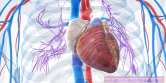
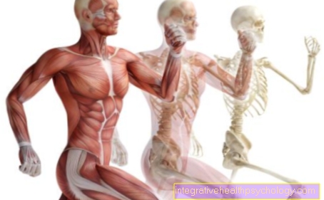
Due to the disturbed and reduced collagen synthesis, the Ehlers-Danlos Syndrome the parts of the body that are particularly rich in connective tissue: the skin, Joints and Blood vessels. Since the connective tissue is lacking in strength, it is overstretchable and tears very quickly, which is especially true for blood vessels that are too small, but sometimes too massive bleeding can lead. An important complication is the formation of bulges in the vessels, so-called. Aneurysms, with the risk of rupture.
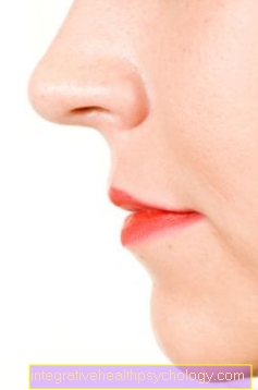
The symptom of the skin is the most pronounced Hyperelastic cutison the side of the neck, over Joints and also in face can be lifted up to 4cm or more. After letting go, it immediately springs back to its starting position, which is why it got the name "Rubber skin" wearing. In general, the skin is noticeably thin (like cigarette paper), soft and velvety ("Marshmallow Skin").
Wounds show delayed wound healing, so that sutures take 3 to 4 times longer to heal. Atrophic or hypertrophic, inferior ones often develop from the seams scar. In addition, liquid-filled (succulent) bulges of the skin (mollusoid pseudotumors) develop on heavily stressed areas of the body, such as Knee and elbow joint, to the formation of ankle pads ("Knuckle pads") on Back of hand and foot and of nodules on the heel.
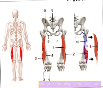
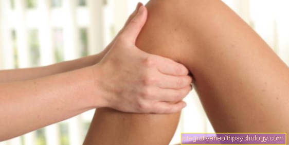
The Joints can be overstretched (Hyperflexibility), often movable in unwanted directions and lacking strength due to loosened joint ligaments (Ligament laxity). This can cause unusual movements, such as those one might expect from "Contortionists" knows. The joints tend to Contortions (Dislocations) and malpositions. Those are particularly affected shoulder- and Ankle joints, the Kneecap (patella), the Temporomandibular joint (Temporo-mandibular joint) and more rarely that Elbow joint. The documentation of overmobility of the joints (hypermobility) is carried out by the Beighton's scorewho confirms hypermobility on 5 out of 9 possible points.
Other symptoms of the joints are generalized joint problems, chronic neck pain, move- and Hip pain, Joint and Muscle achesthat are difficult to treat. Sometimes pain points ("Tender ponits"), which are defined as an area that reacts painfully to pressure loads of 4 kg or less. In addition, there is an increased risk of fractures due to reduced bone mass combined with an abnormal bone structure.
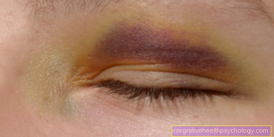
Due to the fragility of the connective tissue of the blood vessels, there is a pronounced tendency to Hematomas, spontaneously or as a result of trauma,
mainly in areas at risk of injury. This is followed by a typical in the affected areas brown pigmentation.
After injuries one observes a tendency to prolonged bleeding with normal coagulation values. The fragility of larger blood vessels can be triggered by exertion, accidents, pregnancy or birth lead to massive, life-threatening bleeding.
Since other connective tissue structures are also inferior, it can become too Viscera (Hernia / inguinal hernia)), curvature of the spine (Scoliosis), Cracks (rupture) of the Intestines and the uterus (Uterus), sacs (Aneurysm) of blood vessels and constriction of the lungs from free air in the chest (Pneumothorax) come.
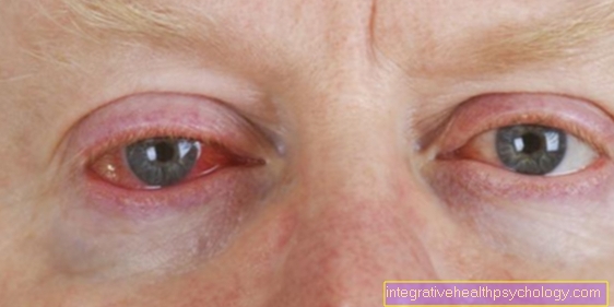
In rare cases, eye changes are associated with EDS such as Astigmatism (astigmatism) or green Star (glaucoma) to observe.
diagnosis
The diagnosis is made on the basis of the clinical appearance, the symptoms, and is supplemented by a family examination (family history). In addition, a Skin biopsy in which the removed skin tissue is examined using an electron microscope and its collagen structure is assessed. The differentiation into the different types of Ehlers-Danlos Syndrome takes place by means of sequence analysis of the DNA.
Classification / types
Type I, II: Classic guy; Inheritance: autosomal dominant; symptoms: Hyperelasticity and fragility of the skin, atrophic scarring, joint hypermobility; Cause: collagen V formation disorder
Type III: Hypermobile type; Inheritance: autosomal dominant; symptoms: generalized joint hypermobility, skin involvement (hyperelasticity and / or soft, vulnerable skin); Cause: collagen V formation disorder
Read more about this important type at: Ehlers-Danlos syndrome type III
Type IV: Vascular type; Inheritance: autosomal dominant; symptoms: thin translucent skin, ruptures of the arteries, intestines and uterus, pronounced tendency to hematoma; Cause: collagen III formation disorder
Type V: corresponds to type I.
Type VI: Kyphoscoliotic type; Inheritance: autosomal recessive; symptoms: decreased tension of the Musculature already at birth ("Floppy infant"), delayed development of holding and supporting reflexes, lateral bending of the Spine (Scoliosis), Root cause: Lsysl hydroxylase deficiency
Type VII A / B: Arthrochalastic type; Inheritance: autosomal dominant; symptoms: severe generalized hypermobility of the joints with repeated dislocations, congenital bilateral hip dislocation; Root cause: Type I collagen disorder
Type VII C: Dermatosparactic type; Inheritance: autosomal dominant; symptoms: pronounced skin fragility, sagging skin, Root cause: Deficiency of N-terminal procollagen I peptidase
Therapy and prophylaxis
Neither one causal still a symptomatic therapy is currently possible, so the prophylaxis of consequential damage is in the foreground. Injuries and greater stress on the joints should be avoided. So should certain sportswhich are associated with an increased risk of injury are not exercised. Due to the increased risk of complications pregnancy and birth both Types I, II, IV and VI close monitoring is required.
It should also be used with colds cough suppressant therapy and generally to the Regulation of stool consistency to be respected, there is such a thing Colon rupture (Colon rupture) and a Pneumothorax can be avoided. Through early physiotherapy, especially in children, the overstretchable joints can be stabilized, which leads to the alleviation of the symptoms of the entire musculoskeletal system.
Wounds special care must be taken and operations should only be carried out in emergencies because wound healing takes 3 to 4 times slower than usual.
forecast
Patient with Ehlers-Danlos Syndrome usually have a normal life expectancy. However, the disease is progressive, so it leads to an ever-increasing deterioration in health. Skin wounds and joint dislocations affect the patient's quality of life, while rupture of the great vessels can be life-threatening.
Life expectancy
Ehlers-Danlos syndrome is a chronic disease for which there is still no causal treatment and therefore no cure. This means that, given the current state of medical technology, there is no way to do anything about the causes of Ehlers-Danlos syndrome and to cure it completely. Unfortunately, one is still not able to combat and treat the symptoms that occur. The only thing you can do is to encourage the affected patient to always be careful not to put too much strain on the joints and to avoid injuries to the skin whenever possible. Surgical interventions should only be carried out in an emergency and if there are no adequate alternatives.
In most cases it is progressive with an increasing worsening of symptoms and impairments in the everyday life of the patient. Depending on the type of disease, the disease has different effects on the lives of those affected. The changes in the joints sometimes lead to osteoarthritis and arthritis in early childhood, so that the children later learn to walk and their feet may be deformed. With the increased risk of retinal detachment or retinal hemorrhage, eyesight is also compromised.
How strongly the symptoms are ultimately pronounced in each individual depends heavily on the type of Ehlers-Danlos syndrome and they can also vary greatly within the individual subtypes. For most types of Ehlers-Danlos syndrome, life expectancy is normal. In patients with Ehlers-Danlos syndrome type IV, which affects the vessels, life expectancy is significantly reduced due to serious complications, such as the risk of spontaneous rupture of an artery, especially the main artery (med. Aortic tear) or the large intestine.
It is around 37 years for women and 34 years for men.
A reduced life expectancy can also be assumed with Ehlers-Danlos syndrome type VI.