Diseases of the knee joint
Here you will find the most important anatomical principles of a healthy knee joint and an overview of all major knee joint diseases with a brief definition of the disease.
For more detailed information, simply follow the link to the appropriate disease.

Classification of diseases of the knee joint
The following is an overview of the most common diseases of the knee joint, sorted by:
- Injuries to the ligaments of the knee joint
- Injuries to the bony structures of the knee joint
- Diseases caused by overuse and wear and tear
- Inflammation in the knee
- Specific diseases of the knee joint
If you are not sure what is causing your knee pain, we recommend our self-test to find out the cause and the treatment options.
Appointment with a knee specialist?

I would be happy to advise you!
Who am I?
My name is I am a specialist in orthopedics and the founder of .
Various television programs and print media report regularly about my work. On HR television you can see me every 6 weeks live on "Hallo Hessen".
But now enough is indicated ;-)
The knee joint is one of the joints with the greatest stress.
Therefore, the treatment of the knee joint (e.g. meniscus tear, cartilage damage, cruciate ligament damage, runner's knee, etc.) requires a lot of experience.
I treat a wide variety of knee diseases in a conservative way.
The aim of any treatment is treatment without surgery.
Which therapy achieves the best results in the long term can only be determined after looking at all of the information (Examination, X-ray, ultrasound, MRI, etc.) be assessed.
You can find me in:
- - your orthopedic surgeon
14
Directly to the online appointment arrangement
Unfortunately, it is currently only possible to make an appointment with private health insurers. I hope for your understanding!
Further information about myself can be found at
Injuries to the ligaments of the knee joint

Meniscus damage
Meniscus damage is understood to be the injury or tear to one of the two cartilage discs that are located between the thigh and shin bones. The joint surfaces of the thigh and shin do not fit together. To compensate for this "asymmetry" we have "cartilage discs" in the joint, an inner and an outer meniscus.
Further information is available under our topic: Meniscus damage
Anterior cruciate ligament tear
A fresh anterior cruciate ligament tear is the complete or partial interruption of continuity (Crack) of the tape after exceeding the overstretch reserve due to external force. An old anterior cruciate ligament rupture is a permanent, mostly accident-related ligament injury. Typically, such an injury can occur when the lower leg is stationary while skiing or playing soccer.
Read more on this topic at: anterior cruciate ligament tear
Posterior cruciate ligament tear
A fresh posterior cruciate ligament rupture is the complete or partial interruption of the continuity (tear) of the ligament after the overstretch reserve has been exceeded by external force. An old posterior cruciate ligament rupture is the permanent, mostly accident-related ligament damage.
Read more on this topic at: posterior cruciate ligament rupture
Outer ligament tear
In the case of lateral ligament injuries, it is usually a tear of the same. Usually the tear is complete - there are hardly any incomplete tape tears. The cause is usually trauma (rotation, dislocation). The therapy depends on the extent of the injury (outer ligament tear), and can range from immobilization for a few days to surgery. The prognosis is usually good.
Read more on this topic at: External ligament tear on the knee
Inner ligament tear
In the case of internal ligament injuries, it is usually a tear of the same. Usually the tear is complete - there are hardly any incomplete tape tears. The inner ligament usually only breaks as a result of trauma. This can be a kinking, a rotational trauma or a knee joint dislocation, such as those that occur when skiing or playing soccer. Therapy for the torn inner ligament depends on the extent of the injury.
Read more on this topic at: Inner ligament tear in the knee
Ligament stretch
A ligament stretch (Syn. Ligament strain) of the knee is caused by excessive movement of the knee joint that is too violent and can affect both the inner and outer ligaments. It is one of the most common sports injuries and can result, for example, from a sudden rotational movement of the knee.
Read more on this topic at: Ligament stretch in the knee
Patellar tendon tear
A patellar tendon rupture occurs when the tendon between the anterior thigh muscles and the lower part of the kneecap tears partially or completely. The term is also synonymous with the patellar tendon tear Patellar tendon rupture used. The patellar tendon rupture usually occurs spontaneously due to excessive tension in the leg against resistance or when the knee is tensed in a flexed position. Those affected usually express a sudden pain.
Read more about this under: Patellar tendon tear
Injuries to the bony structures of the knee

Kneecap fracture
When the kneecap breaks, the kneecap fractures into several parts. This can result in longitudinal, transverse or mixed breaks. The treatment of the kneecap / patellar fracture depends crucially on the type of fracture. A doctor should be consulted for further diagnosis.
Further information is available under our topic: Patellar fracture
Shin splints
Shin splint syndrome is a chronic pain syndrome in the area of one or more fascial compartments of the lower leg due to disproportions between muscles and fascia that arise during exercise. The main symptoms of tibial splint syndrome are primarily pain, initially when moving, later also possible at rest. When treating shin splints, a distinction must be made between conservative and surgical treatment.
Read more on this topic at: Shin splints
Bruised tibia
Reasons for a bruised shinbone are very straightforward. A bruised shin bone is usually contracted by hitting the shin or kicking a rigid or solid object that cannot yield. The classic symptoms of a bruised shinbone are pain, swelling, a bruise, and some restriction of movement.
Read more on this topic at: Bruised tibia
Diseases due to overuse and wear and tear
Cartilage damage in the knee
Cartilage damage in the knee is quite common. Most of the time, the damage is caused by wear and tear. On the one hand, this wear and tear occurs as part of a completely natural aging process. The expert calls the result of this arthritis. First of all, it is important that all risk factors are eliminated or reduced as far as possible.
Read more on this topic at: Cartilage damage in the knee
Jumper knees
The jumper's knee is a chronic, painful, degenerative disorder of the kneecap extensor system at the bone / tendon junction of the kneecap tip. It is also known as "patellar tip syndrome". Patients report stress-dependent pain in the area of the kneecap tip.
Further information is available under our topic: Jumper knees
Osteoarthritis of the knee
Under Gonarthrosis / knee joint arthrosis All degenerative (wear-related) diseases of the knee joint are to be understood, which are characterized by an increasing destruction of the articular cartilage with the involvement of the joint structures such as bones, joint capsules and muscles near the joint. It can have several causes and usually shows itself initially as a start-up pain and swelling.
Further information is available under our topic: Gonarthrosis / knee joint arthrosis
Kneecap dislocation
In the typical patellar dislocation, the kneecap jumps out of the intended slideway. This often leads to ligament, cartilage and bone injuries. The symptoms of a kneecap dislocation (kneecap dislocation) are usually so typical that they allow the experienced doctor to make a visual diagnosis. In most cases, a dislocation of the kneecap does not require any treatment, as it usually springs back into its plain bearing by itself
Read more about this under: Patellar luxation
Shelf Syndrome
Shelf syndrome occurs after overuse, muscle imbalance or injury to the knee. It is caused by inflammation and swelling of the folds of the mucous membrane (synovial folds, plicae) in the knee joint. This can lead to pain and restricted mobility in the knee joint.
Three folds of the mucous membrane of the knee can be affected: the plica suprapatellaris, the plica mediopatellaris, and the plica infrapatellaris. By far the most common is the mediopatellar plica affected.
Read more on this topic at: Shelf Syndrome
Inflammation in the knee

Inflammation of the kneecap
Like almost any other organ, the kneecap can become inflamed. This can lead to considerable pain and restricted mobility in the knee joint. The main symptom of inflammation of the knee joint is knee pain, especially in the front area of the knee joint and directly above / below the kneecap. The most common cause of inflammation of the kneecap is overload.
Read more on this topic at: Inflammation of the kneecap
Inflammation of the patellar tendon
Incorrect or excessive strain on the knee can easily lead to irritation and inflammation of the patellar tendon, which is associated with severe knee pain and restricted mobility. Pain at the lower end of the kneecap is typical. The most important measure in therapy for inflammation of the patellar tendon is protection.
Read more on this topic at: Inflammation of the patellar tendon
Inflammation of the quadriceps tendon
In most cases, inflammation of the quadriceps tendon is caused by chronic overloading of the tendon and all of the structures connected to it. The person concerned becomes aware of it primarily through a punctiform tenderness exactly over the corresponding tendon section. To treat inflammation of the quadriceps tendon, conservative therapy can be considered at the beginning.
Read more on this topic at: Inflammation of the quadriceps tendon
Specific diseases of the knee joint

Ahlbäck's disease
The term Ahlbäck M. The reasons for the occurrence of M. Ahlbäck are based in most cases on an inferior blood flow to the lower part of the thigh.
Read more on this topic at: Ahlbäck's disease
Sinding-Larsen's disease
The disease known as Sinding-Larsen's disease is an extremely painful inflammatory reaction in the knee joint. The majority of affected patients are young people. Sinding-Larsen's disease is more common in athletes. The treatment of Sinding-Larsen's disease is divided into non-operative and operative measures.
Read more on this topic at: Sinding-Larsen's disease
Arthrofibrosis
Arthrofibrosis is a dreaded joint disease, largely unexplained in its cause, after surgery or injuries, which results in a more or less severe, sometimes painful, limitation of joint mobility. A distinction is made between primary arthrofibrosis, which is characterized by generalized scarring in the joint, and secondary arthrofibrosis, in which local mechanical irritators are the cause of restricted mobility.
Further information is available under our topic: Arthrofibrosis
Osgood - Schlatter disease
Osgood-Schlatter's disease is a painful irritation of the insertion of the kneecap tendon (patellar tendon) on the anterior tibia. This can lead to a detachment and death (necrosis) of pieces of bone from the shin. A dead bone area is created. Since this osteonecrosis is not infectious (not caused by bacteria, viruses or any other), it belongs to the group of aseptic osteonecrosis.
Further information on this topic is available at: Osgood-Schlatter disease
Baker's cyst
A Baker cyst is a protrusion of the posterior joint capsule caused by internal knee disease with chronic knee joint effusion. A Baker's cyst is particularly common in older people due to wear and tear of the knee joint and in children (usually without a clear cause). The diagnosis of Baker's cyst can generally be made by a doctor.
Further information is available under our topic: Baker's cyst
Osteochondrosis dissecans
Osteochondrosis dissecans (OD) is a disease that often occurs in growing age and young adulthood, about 85% of which affects the knee joint. In the course of this disease, bone death near the cartilage occurs, whereby a piece of cartilage located above the affected bone area can become detached from its composite.
Further information is available under our topic: Osteochondrosis dissecans
Patellofemoral pain syndrome
The pain of the kneecap is also known as femoropatellar pain syndrome (PFFS). PFFS is one of the most common symptoms in the anterior knee area. The PFSS does not hide a uniform clinical picture, but rather a very complex symptom picture, which is discussed very differently in terms of definition, diagnosis and etiology (causes).
Read more on this topic at: Patellofemoral pain syndrome
General information about the knee joint
Here you can find more information about the structure and function of the knee joint and common symptoms.
The anatomical structure of the knee joint
The anatomy of the healthy knee joint
The knee joint is the largest Joint of man and provides the flexible connection between Thigh (Femur) and Lower leg (Tibia). Three bone together with a complex capsular and ligamentous system (Collateral and cruciate ligaments) the framework of the knee joint.
These are:
- the Thigh rolls (Femoral condyles)
- of the Tibial head (Tibial plateau)
- the Kneecap (patella)
Illustration of the knee joint

A - Right knee joint from the left
B - Right knee joint from the front
C - Right knee joint from behind
- Kneecap - patella
- Femur - Femur
- Shin - Tibia
- Fibula - Fibula
- Inner meniscus -
Meniscus medialis - Outer meniscus -
Lateral meniscus - Kneecap ligament -
Ligamentum patellae - Outer band -
Ligament collaterale fibulare - Inner band -
Ligament collateral tibial - Posterior cruciate ligament -
Ligament cruciatum posterius - Anterior cruciate ligament -
Ligament cruciatum anterius
You can find an overview of all Dr-Gumpert images at: medical illustrations
The figure above shows that the bone have close contact with each other. So that the knee joint can move painlessly and undisturbed on the contact surfaces, the bones on the respective contact surfaces are covered with a very smooth, whitish layer of cartilage, the so-called hyaline cartilage, overdrawn.
Only through them is pain-free and undisturbed mobility of the knee joint possible. The hyaline cartilage is composed of cartilage cells and matrix tissue. While the adult cartilage cells have lost the ability to divide and thus heal, cartilage cells in children can repair cartilage wounds by multiplying.
The cartilage cells are embedded in the matrix tissue they produce themselves. This consists largely of water and supporting tissue and gives the cartilage tissue both stability and elasticity.
in the Knee joint even one differentiates between the joint pairing Thigh roll and the Kneecap (Femoropatellar joint) as well as the thigh roll and the tibia head. The connection between the thigh roll and the kneecap (patella) is the most stressed part of the knee joint. When climbing stairs, for example, this joint becomes more than three times the Body weight burdened.
The most important tendon of the knee joint is the Kneecap tendon (patellar tendon / patellar tendon)running from the kneecap at the front of the knee joint as a strong cord to the shin (Tuberisitas tibiae) pulls.
The knee joint is enveloped by the knee joint capsule, the inner part of which is the synovial membrane (Synovia) referred to as. It is important for the nutrition of the cartilage Synovial fluid. In adulthood, the cartilage tissue is mainly nourished by diffusion (passive transport) from the synovial fluid because, in contrast to other types of tissue in the body (Muscles, bones, skin etc..) is not supplied with blood.
The usual diet via the bloodstream therefore does not work.
Movements of the knee joint mix the synovial fluid and thereby improve the absorption of nutrients by the cartilage cells (Chondrocyte).
The right amount and composition of the synovial fluid is also of crucial importance for the lubrication of the knee joint. They minimize the friction of the corresponding cartilage surfaces during movement. Hyaline cartilage has an extremely low frictional resistance, lower than all known artificial material combinations.
Between the thigh roll and the lower leg there are two cartilaginous disks (meniscus), which act as a buffer between the cartilage surfaces and are important for an even pressure distribution in the knee joint when under stress. The cruciate ligaments run through the knee joint and connect the thigh to the lower leg. The main task of the cruciate ligaments is to ensure a stable knee joint and thereby enable a harmonious, low-friction movement sequence.
The two cruciate ligaments prevent the knee from shifting forwards (anterior cruciate ligament) or back (posterior cruciate ligament) when under stress. In their stabilizing effect, they are supported by the collateral ligaments, the knee joint capsule and the muscles surrounding the knee.
The knee-spanning muscles make up the function of the knee joint. Movement is only possible through them. In addition, they support the passive joint stabilizers of the capsular ligament apparatus. The extensor muscles of the knee joint (quadriceps muscles) on the front of the thigh are very strong. The muscles are involuntarily activated when the knee is bent in order to avoid buckling while walking. The most important flexors of the knee joint are the hamstring muscles on the back of the thigh, which also have an active stabilizing effect on the knee joint. The hamstring muscles support the anterior cruciate ligament in particular, which is why their training after a cruciate ligament rupture is particularly important.
Knee pain
Knee pain can be divided according to where on the knee they occur.
Knee pain on the inside may indicate a lesion of the medial meniscus or the medial ligament. In addition, they often occur in the context of wear and tear, for example in a Osteoarthritis of the knee on the inside of the knee joint. An incorrect load when running due to unsuitable footwear, misalignment of the feet or leg axis as well as a leg length difference can cause pain on the inside of the knee.
The same applies to the outside of the knee joint. Localized pain (please refer: external knee pain), in contrast to the pain on the inside, indicate involvement of the outer ligament or the outer meniscus.
Knee arthrosis can also be the cause of this if it affects the outer knee joint area.In the case of acute injuries to the menisci or ligaments, the pain is usually stabbing, suddenly occurring and is often accompanied by other symptoms such as swelling or overheating of the joint region and joint effusion.
In degenerative processes, such as osteoarthritis, the pain usually develops over a longer period of time and occurs predominantly when the joint is stressed.
Knee pain, predominantly in the hollow of the knee (please refer: Back of the knee pain) focus can also have different causes. An injury to the back of a meniscus can cause pain in the hollow of the knee. Inflammation or changes in muscle tendons and knee arthrosis in this area can also explain the symptoms.
With osteoarthritis or one Meniscus damage Over time, a so-called Baker's cyst come. This is a bulge in the joint capsule that is filled with fluid. From a certain size the Baker cyst can be felt as a swelling in the hollow of the knee and can also cause pain there.
If you have pain in the hollow of your knee, you always have to contact one thrombosis - a blood clot - in the blood vessels of the lower leg.
Knee pain can also be localized behind the kneecap. This is typical of what is known as retropatellar osteoarthritis, an arthritis that occurs directly behind the kneecap (patella) arises. A muscular imbalance or a congenital deformation of the kneecap can also cause pain behind the kneecap, since it cannot slide in the bone groove intended for it. There is friction and wear in the joint and secondary pain.
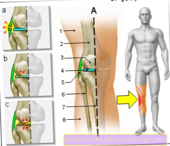
Figure knee pain on the inside

A - Right knee joint from the front
- Inner thigh muscle -
Vastus medialis muscle - Femur -
Femur - Kneecap -
patella - Inner meniscus -
Meniscus medialis - Inner band -
Ligamentum collaterale tibiale - Internal calf muscle
Gastrocnemius muscle,
Caput mediale - Shin -
Tibia
a - Inner ligament injury
(Inner ligament tear)
b - tear of the medial meniscus
c - medial
(internal) knee osteoarthritis
You can find an overview of all Dr-Gumpert images at: medical illustrations
Figure knee pain on the outside

A - Right knee joint from the front
- External hamstring muscle -
Vastus lateralis muscle - Femur -
Femur - Kneecap -
patella - Outer meniscus -
Lateral meniscus - Outer band -
Ligamentum collaterale fibulare - Fibula -
Fibula - Shin -
Tibia - External calf muscle
Gastrocnemius muscle,
Caput laterale
a - Ligament injury
(Outer ligament tear)
b - External meniscus tear
c - Lateral
(external) knee arthrosis
You can find an overview of all Dr-Gumpert images at: medical illustrations
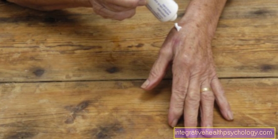
Tap your knees
To stabilize the knee, it can be taped with special tapes.
These so-called Kinesio tapes can be used in many ways and achieve a good effect. In order to optimally relieve and stabilize the knee, the right technique is essential when taping.
To do this, a Y-shaped incised tape stuck on above the kneecap and then the two Y-legs outside and inside around the kneecap so that they meet again below the kneecap. The knee should be bent when gluing. As an alternative to the Y-shaped cutting of the tape, two individual tape strips can be placed around the kneecap in an arc on the outside and inside.
When gluing below the kneecap, no tension should be exerted on the tape. If the leg is now straightened, the skin over the knee should wrinkle. The kneecap is then optimally stabilized in its position and a dislocation under load is prevented.
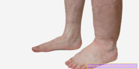
Knees twisted
A twisted knee causes severe pain immediately after the trauma.
Due to the unnatural dislocation, the ligaments in the joint area are overstretched. Blood vessels can tear and bruises can form in the skin and joint effusions.
This often manifests itself as a swelling of the joint. The joint capsule can also be damaged when twisted. The exact extent of the injury can usually not be determined from the clinical picture alone. This requires more precise diagnostic measures.
The recommended acute therapy for the twisted knee joint is Elevation and coolingto prevent further swelling.
If there are no other injuries in or on the joint beyond the strain, no special therapy is required. The pain usually goes away on its own within a few days.
Painkillers can be taken to bridge the gap, as the twisted knee can still cause pain over a long period of time when it is exerted. If ligaments or joint capsules have been more severely damaged, a more detailed diagnosis must be carried out.
In the case of a twisted knee with swelling and / or instability, diagnosis is usually carried out using a MRI of the knee. In the knee MRI, soft tissue damage, such as occurs particularly in the capsule and ligaments, can be particularly well detected.
You can find much more information about what you can and should do under our topic: Knees twisted