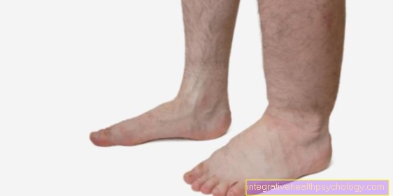
Tarsus
anatomy
The tarsus includes all structures that are between the fibula, shin, and toes. This includes 7 tarsal bones, which can be divided into two rows, but also several joints, as well as the entire ligament and muscular system in this region.

The Tarsus can be divided into one closer to the fuselage, the so-called "proximal"And a"distal“Row of bones. The proximal row consists of the talus "Talus"And the distal bone"Calcaneus". The distal row contains several small bones. These include the navicular bone "Navicular bone", The cuboid"Os cuboideum"And three cuneiform bones"Ossa cuneiform“, Which are again subdivided into a middle, an outer and something in between. The Calcaneus is probably the most prominent, as it forms the classic heel, represents the largest bone in the foot and has to support a large part of the entire body weight.
Read more on the topic: Metatarsal pain.
All Tarsus are closely connected with each other by tight ligaments, which primarily stabilize the two ankle joints and enable an upright, secure stance. The protrusion formed by the calcaneus is also known as "Calcaneal tuberosity". Among other things, it has a function as a starting point for Achilles tendon, which is why this region is known as the Achilles' heel. The Achilles tendon itself is the insertion of a multi-part muscle that arises from the thigh and most of the muscular calf forms. Its function is to straighten the foot as a whole.
The two are primarily responsible for the movements of the foot Hocks significant. Due to the high weight load and stability requirements, the joints are very firmly secured by tapes, which accordingly also limits the mobility compared to the hand. in the upper ankle is the talus "Talus“From above and from left and right completely from the two lower leg bones, the Fibula „Fibula“Outside and that Shin „Tibia“, Enclosed in the middle. This becomes palpable and also visible from the outside in the form of the two ankle. As a result, the main movement in this joint is only the extension or the tightening of the foot ("Extension" and "Flexion"). Different ligaments stretch from bone to bone inside and outside, which have a lateral stabilization function. In summary, these are only called the inner band "Medial collateral ligament" and Outer band "Ligamentum collaterale laterale".
The lower ankle is divided again into one front lower and a rear lower Ankle joint. In the back lower ankle stand that Calcaneus and the Ankle bone articulated with one another, whereas in the anterior lower ankle joint the proximal tarsal bones with the Scaphoid form a joint. Since this joint is about several bones extends and anatomically in two single joint capsules is structured, this must also be done by a multitude tighter ligaments attached and stabilized. The lower ankle joint allows, to a limited extent, the execution of "Supination and pronation movements". That means that you have the Lift the middle and outer edge of the foot can.
Figure ankle

- Toe phalanx - Phalanx distalis
- Middle toe phalanx - Phalanx media
- Metatarsal phalanx - Phal. proximalis
(1st - 3rd toe bones - Phalanges) - Metatarsal bones -
Os metatarsi - Inner sphenoid bone -
Medial cuneiform bone - Middle sphenoid bone -
Os cuneiform intermedium - External sphenoid bone -
Os cuneiform laterale - Cuboid - Os cuboideum
- Scaphoid bone - Navicular bone
- Ankle bone - Talus
- Ankle roll - Trochlea tali
- Heel bone - Calcaneus
- Protrusion on 5th metatarsal - Tuberositas ossis metatarsalis quinti (V)
You can find an overview of all Dr-Gumpert images at: medical illustrations
Tarsus fracture
With the multitude of existing ones Tarsus fractures can occur under certain conditions. Such a break can be differentiated on the basis of various criteria. If there is a break, it divides by definition a contiguous single bone in at least two parts. Such a break almost always goes along with it Pain and one Function restriction hand in hand. Other aspects can play a role in the assessment, for example the root cause the fracture, that extent and the location of the fracture, or whether there is an open fracture (whether an open wound has occurred due to bone parts).
The two most common causes of a fracture of the Tarsus are an action of force, for example in the course of a Accident, and a metabolic one Weakening of the bonesas they are, for example, with age-related osteoporosis can occur, which means that normal (physiological) stress already leads to a fracture. Such a fracture can show up through being visible abnormal positions the bone. Otherwise it can be assumed in the case of typical locally limited ones Pain, especially when performing movements of the involved bone, as well as when Swelling, tenderness, or bruising. When describing the course of the accident and these symptoms, a X-ray image recorded in the affected region. At the tarsus in particular, it is essential to scan several anatomical levels of the foot, since often not all bones and fractures can only be seen from one angle. In addition, a higher-resolution image can be provided with a CT or MRI provide more information.
If such a fracture has been diagnosed, the doctor will decide on further therapeutic measures depending on the position of the bone fragments. Surgery may be required. The goal here is to bring the bone fragments inside the tarsus to their original place and to connect them there with wires or screws. Complete healing of the bone parts takes a few weeks. In the meantime, it is important to relieve the fracture site and immobilize it, while at the same time maintaining and exercising the function of the joints and muscles. A plaster cast or splint is often required to relieve the strain, usually in combination with walking aids.
Read more on this topic at: Broken foot.
injury
Due to the high weight load that our feet are physiologically exposed to every day, they are predestined for injuries and trauma caused by an accident.
In addition to the tarsus fractures described above, the "twisting trauma" is a common injury. The classic twisting of the foot inwards or outwards can occur at any time in everyday life or frequently in many sports. Particularly leg-heavy and jump-heavy sports are high risk factors, for example soccer, basketball or handball. If it happens, you should immediately relieve the foot. At the same time, you can start to cool the affected area, raise it up and press it lightly in order to counteract any swelling that is beginning to occur. In any event, a doctor should be consulted to assess the extent of the injury. This uses ultrasound, an X-ray image, CT or MRT. The MRI in particular can provide precise information about the damage to the tissue or the involvement of the bone.
Read more on the topic: Foot bend over.
Often there is a strain or tear in one of the tight ligaments of the upper ankle, the "Collateral ligaments". The outer ligament on the foot is particularly often affected, as a so-called "Supination trauma", An outward twisting, is much more common than a"Pronation trauma". In most cases, however, treatment is conservative by protecting the area, often supplemented by bandages or splints. Under certain circumstances, however, an operative supplement may be necessary, for example if the connection between the two bones of the lower leg, the "Syndesmosis“, Is injured. In many cases, such an injury subsides without consequences. However, there is also the risk of permanent instability remaining, which means that twisting an ankle can become more and more common.
You might also be interested in: Achilles heel