Xanthelasma
Definition of xanthelasma
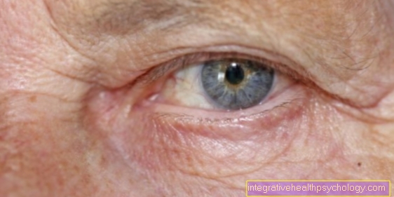
Xanthelasma are yellowish plaques that are created by lipid deposits (lipids are fats, especially cholesterol) in the upper and lower eyelids. They are harmless, in no case contagious and also not hereditary, although they can occur in families.

When do xanthelasma occur?
Xanthelasma can appear at any age, but are most common between the ages of 40 and 60 when they first appear.
Women are more often affected than men, but an unhealthy lifestyle, smoking, fatty foods and obesity, as well as a predisposition to high cholesterol are risk factors for xanthelasma and subsequent stroke or heart attack.
Summary
In this clinical picture, lipids are stored in the skin the upper and / or lower eyelids. Lipids are fats. In older people this often happens without a cause, in younger people underlying diseases must be ruled out. One recognizes the xanthelasma as yellowish cushions. If desired, cut out the affected areas of the skin.
Recognizing xanthelasma
What are the symptoms of xanthelasma?
Most xanthelasma are located in the area of the inner corner of the eye. The upper eyelid is affected more often than the lower eyelid.
The xanthelasma are striking due to their raised surface and yellow skin discoloration. Xanthelasma are soft and movable.
The course is very different: From long-term constant courses to the size and distribution of increasing xanthelasma, everything was observed.
Xanthelasma is not painful and does not cause any other complaints, but those affected or relatives usually notice it as a cosmetic impairment. In rare cases, the function of the eyelids can be restricted, so that the eyelid on the affected side hangs down more (ptosis).
With the corresponding underlying disease, symptoms can also occur that are not caused by the xanthelasma. Such fat deposits in cells also occur in other parts of the body, e.g. E.g. tendons, before.
How are xanthelasma diagnosed?
The diagnosis of xanthelasma is a visual diagnosis, because the xanthelasma can be seen with the naked eye.
For younger people, additional diagnostics, such as E.g. blood samples to determine blood values, run to prevent an underlying disease (Hyperlipidemia) to be excluded.
Treating xanthelasma
How are xanthelasma treated?
Conservative:
If there is a disorder in lipid metabolism, treatment of the underlying disease with lipid-lowering drugs and diet is indicated so that the progression of the metabolic disorder and its numerous consequences are prevented.
However, diet and lipid-lowering therapy can usually only have little influence on the xanthelasma.
Surgical:
To remove xanthelasma, these are surgically removed. There are different methods: excision, cauterization with HF devices or chloroacetic acid.
Today the procedure is mainly performed using laser ablation and CO2 lasers. Since the reason for the removal is mostly cosmetic damage, the statutory health insurance does not pay for the procedure.
Regardless of which surgical method is chosen, care must be taken as the eyelid has a very special anatomy. If too much tissue is removed, the subsequent shrinkage of the scars can lead to eyelid closure disorders (ectropion), so that the surface of the eye (cornea) can dry out. Pigment disorders are also a risk of complications that would lead to an aesthetically unsatisfactory surgical result.
Read more on the topic: Xanthelasma surgery and Remove xanthelasma
Can xanthelasma be operated on?
Surgical removal of xanthelasma becomes necessary if the patient suffers too much from the cosmetic impairment caused by the xanthelasma or if their position and size hinder the eyelid closure. The xanthelasma themselves are benign and so do not necessarily have to be removed.
The intervention is a quick routine matter and can be performed on an outpatient basis and under local anesthesia. The doctor either traditionally chooses the scalpel or a laser, which, however, makes the treatment more expensive and complex without offering any cosmetic advantage. The affected skin area is cut out with a scalpel and the eyelid is then tightened. The operation is therefore not always possible, as there must be enough skin to close the wound. The skin of the eyelids also tends to recur. In 40% of the cases, new xanthelasma form in the same places after the removal, after the second operation it is already 60%. Whichever type of surgical removal you choose, you have to bear the costs yourself in any case, as the procedure falls under cosmetic treatments. You have to calculate about 250 €, depending on the size and number of xanthelasma and the type of therapy.
Prevention of xanthelasma
What are the causes of xanthelasma?
In the elderly, xanthelasma often occurs for no apparent reason. However, if such lipids are found in the eyelid membranes of younger people, a further investigation should be carried out.
The reason is probably hyperlipidemia (hyper = (too) much; lipids = fats). The affected patients have dissolved too many fats in their blood. Normally, fats in the blood are absorbed by the responsible cells and transported to the liver to be metabolized there. The xanthelasma is therefore a disorder in the fat metabolism, as a result of which the body stores the excess fat in the area around the eyes.
The body has excess fat either because it absorbs too much fat when digesting food or because it is not processing the fat properly.
In around 50% of those affected, lipid metabolic disorders such as diagnose type II or type IV hyperlipidemia. This lipid metabolism disorder is often associated with diabetes mellitus.
Xanthelasma can also occur in people with normal total cholesterol values but low HDL levels.
Read more about the topic here: Lipid metabolism disorder
It is important for those affected to have an extended check-up at the family doctor, who can clarify an increased risk of cardiovascular diseases. During the examination, blood pressure should be measured, as well as weight and waist circumference, and a blood test for cholesterol and triglyceride levels should be carried out. Furthermore, advanced technical procedures (e.g. ultrasound) can be used to examine the blood vessels for an existing disease.
A possibly present arteriosclerosis (narrowing of the blood vessels by deposits) can be diagnosed. This arteriosclerosis can lead to strokes and heart attacks and should be monitored regularly and supported by medication.
Read more on the topic: Xanthelasma causes
Course of xanthelasma
What is the prognosis for xanthelasma?
If the xanthelasma is not completely removed, they can come back. Otherwise there is no danger from the lipia deposits.
Further questions about xanthelasma
Should one express xanthelasma?
When it comes to so-called hard xanthelasma, it is sometimes possible to scratch and, so to speak, express it in the course of a small surgical operation. However, it is not possible to express the xanthelasma yourself, as you would for example with conventional pimples. This is because, unlike pimples, xanthelasma is fatty, chronic deposits and not an acute inflammatory process with pus formation. Therefore, those affected should keep their hands off the xanthelasma and refrain from tampering with them, but rather consult a specialist in skin diseases. They can offer professional help.
Why do xanthelasma occur after pregnancy?
Xanthelasma is caused by an excess of blood lipids in the body. The body cannot store them in any other way and forms small pads on the eyelids. Pregnancy, which in hormonal terms means an enormous change for the expectant mother, can lead to fluctuations and malfunctions in the metabolism, which also has an impact on cholesterol formation. If xanthelasma reoccurs during pregnancy or afterwards, the person affected should consult a family doctor and have them investigate possible causes. It could be, for example, that (pregnancy) diabetes has developed or an underactive thyroid. Both could result in xanthelasma. Since these are in any case of a benign nature and primarily disturb from a cosmetic point of view, the patient does not have to worry about the consequences for her or her child.
How are xanthelasma structured?
The xanthelasma are composed of so-called xanthoma or foam cells. These are histiocytes (macrophages, scavenger cells) that have a “foamy” cytoplasm due to the intracellular storage of fats (lipids). The composition of these lipids has a very high cholesterol content.


