Inflammation of the gallbladder
Synonyms in a broader sense
Cholecystitis, bile, gallbladder, gallstones, cholelithiasis, cholangitis, pancreatitis
Definition and introduction
In the case of inflammation of the gallbladder / Cholel cystitis it is an inflammation of the gallbladder.
Gallstone disease is the most common cause of illness. When gallstones start to move, they often get stuck in narrow spaces and lead to symptoms such as pain, accumulation of secretion and inflammation.
A gallstone disease is called Cholelithiasis designated.
If the stones are in the gallbladder, one speaks of a Cholecystolithiasisif they are in the main bile duct one speaks of Choledocholithiasis.
The treatment of choice for gallbladder inflammation is minimally invasive surgical removal of the gallbladder. In industrialized countries, gallstones are seen as a result of prosperity with malnutrition, obesity, lack of exercise and stress. However, the different incidence in different ethnic groups and an increased family occurrence speak in favor of genetic involvement.
There are also some favorable risk factors.
These are easy to remember via the 6 F's:
- Fat (overweight)
- Female
- Fertile (fertility)
- Forty (> 40 years of age)
- Fair (light skin type)
and - Family (gallbladder infections in the family).
Classification of gallbladder inflammation
- acute
- chronic
- akalkulös (stone-free)
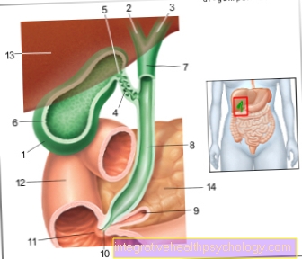
Illustration of the gallbladder

- Gallbladder Body -
Corpus vesicae biliaris - Right liver bile duct -
Ductus hepaticus dexter - Left liver bile duct -
Left hepatic duct - Gallbladder duct -
Cystic duct - Gallbladder Neck -
Collum vesicae biliaris - Mucous membrane -Tunica musoca
- Common
Liver bile duct -
Common hepatic duct - bile duct -
Common bile duct - Pancreatic duct -
Pancreatic duct - Extension of the united
Execution corridor -
Ampula hepatopancreatica - Large duodenal papilla -
Major duodenal papilla - Duodenum Descending Part -
Duodenum, descending part - Liver, diaphragmatic side -
Hepar, Facies diaphragmatica - Pancreas -
Pancreas
You can find an overview of all Dr-Gumpert images at: medical illustrations
Causes of inflammation of the gallbladder

Gallstones - the most common cause of gallbladder inflammation
The causes of acute gallbladder inflammation can be different. However, in 95% of the cases it is a gallstone disease (Cholecystolithiasis). By relocating (obstruction) of the gallbladder duct (Cystic duct) by gallstones, in addition to a build-up of the bile, the mucous membrane of the duct is damaged and an inflammatory reaction occurs. In half of the cases, pathogens such as E. coli, enterococci, salmonella, klebsillia, clostridia etc. a secondary bacterial infection.
Inflammation of the gallbladder without the involvement of gallstones
As already mentioned with the causes, gallstones are the most common cause a gallbladder infection gallstones. Another form of Inflammation of the gallbladder denotes the stoneless (acalculent) Cholecystitis.
Circa 5- 10% to step without the involvement of gallstones on. Gallbladder infections without stones are called acalculous cholecystitis designated.
The cause is a "stress gall bladder". This includes big ones surgical interventions (especially abdominal surgery), Accidents with multiple injuries (Multiple trauma), severe burns, systemic infections (Sepsis) or a impaired blood supply due to vascular diseases (e.g. Polyarteritis nodosa, a Autoimmune disease). This leads to a functional closure of the bile duct and thus to bile congestion and thickening.
A chronic inflammation in most cases results from recurring, recurring acute gallbladder infections.
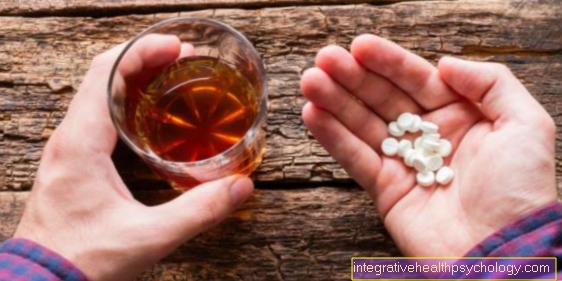
Very rare gallbladder infection poisonous substances or. Medication or causing too large meals.
Epidemiology / frequency distribution

The most common cause of one Inflammation of the gallbladder is a stone disease (see also: Gallstones). Both sexes are affected, but women are about twice as likely to develop gallstones as men. Therefore, the rate of inflammation is also higher in women. Gallbladder infections can occur in all age groups. Around 10-15% of the adult population suffers from gallstones, usually without symptoms for many years. Industrialized countries show a high incidence rate. Some Indian groups are even more likely to suffer from it, while African Americans, East Asians and South Africans are less likely to suffer from it. This suggests a genetic component in the development of gallstones and thus gallbladder inflammation.
Symptoms of gallbladder infection
Pain, nausea, vomiting:
An acute gallbladder inflammation initially manifests itself in severe persistent pain in the right upper abdomen, which often radiates into the shoulder. The pain is often accompanied by nausea and nausea.
Read more on the topic: Gallbladder pain, upper abdominal pain, or right flank pain
Fever:
Persistent fever for 24 hours and an aversion to certain foods are other symptoms of gallbladder infection. Often foods that tend to be fatty and / or promote bile production are avoided (e.g. coffee).
Yellowing:
It is not uncommon for those affected with gallbladder inflammation to have yellow skin (post-hepatic jaundice). Since the bile cannot drain away, certain substances that are normally excreted with the bile via the intestine cannot drain away. Bilirubin, a breakdown product of the blood pigment and oxygen transporter hemoglobin in the red blood cells (erythrocytes), can no longer be properly disposed of from the blood via the liver and bile into the intestine. At first only elevated blood values are shown (Hyperbilirubinemia). If a critical threshold of the concentration is exceeded, the bilirubin escapes from the blood vessels and is deposited in the tissue, which occurs on the body surface or in the dermis (Sclera) of the eyes appears yellow. The urine is also brown in color. The stool, on the other hand, is discolored because it is not excreted via the intestines. In addition, there are fatty stools (Steatorrhea). Fatty stools arise when the bile secretion does not emulsify the dietary fats in the small intestine and so the fats have to be excreted again via the intestine.
Also read: Colors of bowel movements
Chronic gallbladder inflammation:
In contrast, chronic inflammation of the gallbladder shows rather unspecific symptoms such as malaise or digestive disorders such as flatulence and nausea. Repeated biliary colic when gallstones migrate is not uncommon.
Diarrhea - a symptom of gallbladder inflammation ?:
Diarrhea is not a typical symptom of gallbladder infections. Nevertheless, diarrhea can occur in some people affected as their general condition deteriorates. Very light and soft stools can be caused by too little bile. In contrast, the urine is often darker than usual. In any case, a doctor should be consulted for further clarification.
Shortly after the gallbladder has been removed, some sufferers develop diarrhea after eating fatty or spicy foods, as there is no longer any storage reservoir for the bile.
Diagnosis of inflammation of the gallbladder
Diagnosis of gallbladder inflammation, also known as cholecystitis various possibilities into consideration.
1. Anamnesis:
First and foremost, of course, is information gathered from anamnestic. The affected person usually complains of pain in the right upper abdomen below the rib. The pain often radiates to the right shoulder, as this area (head zone) is connected to the gallbladder via nerves. The pain occurs mainly after eating fatty foods or at night. If there is a build-up of bile, the affected person's skin appears yellowish (icteric). Another clue for the inflammation is a fever.
2. Physical examination:
A typical Murphy sign shows up on physical exam. The doctor puts his hand under the patient's right costal arch while the patient inhales. The gall bladder moves downwards and can be felt. If inhalation is stopped due to pain, the Murphy sign is positive. In addition, the person concerned shows a pain-related defensive tension in the abdominal wall.
3. Ultrasound of the abdomen:
For further diagnosis, an ultrasound of the abdomen should be arranged. Gallbladder stones or sludge can be discovered as the cause of the inflammation. If there is cholecystitis, the gallbladder wall is thickened and shows a typical three-layer structure. The gallbladder itself also appears enlarged and can be surrounded by a dark rim of liquid. If free fluid is found, the gallbladder has ruptured. This is an emergency that requires immediate surgery.
Read more on the topic: Ultrasound of the abdomen
4. Blood count:
Gallbladder inflammation can be recognized in the blood based on various inflammation parameters. The main result is an increase in white blood cells (leukocytosis). In addition, the C-reactive protein (CRP) and the erythrocyte sedimentation rate (ESR) are increased.
Some liver enzymes called transaminases, such as GOT and GPT, can also increase. There are also cholestasis parameters that increase when bile builds up. These include the alkaline phosphatase AP, y-GT and direct bilirubin.
Read more on the topic: Gallbladder Inflammation Diagnosis
Therapy for gallbladder inflammation
Surgical therapy for gallbladder inflammation
The therapy for gallbladder inflammation is used nowadays operational by default carried out. If the inflammation is mild, the operation should be performed within the first three days after the onset of symptoms respectively.
In the past, one usually waited up to 6 weeks and only operated on when the patient was symptom-free again. However, studies have shown that prompt surgery brings a better outcome for the patient.
There will also be symptomatic gallstone carriers only operated on. An operation is necessary even at proven stone disease to the exclusion of other causes.
The gallbladder is filled with stones that may be in it completely removed. One speaks of a cholecystectomy. The intervention takes place laparoscopicas this is the case for the patient gentlest procedure is. It just becomes small abdominal cuts needed to introduce the instruments. This is how not a large surgical scar.
If the gallbladder cannot be removed in this way, for example because the stones are too large or there are too many adhesions in the abdominal cavity, a must nevertheless Costal margin be set. This procedure is called an open cholecystectomy.
Multimorbid patients who have a high surgical risk have e.g. due to old age or certain previous illnesses, may initially conservative be treated, including the Administration of antibiotics, such as Anti-inflammatory agents (Anti-inflammatory drugs) and Means against a convulsive and painful contraction of the gallbladder (Antispasmodics and analgesics).
If possible, it should be a surgery follow to avoid complications of cholecystitis. Alternatively can Bile fluid punctured and drained using CT become. This is called percutaneous drainage.
Gallstones, which are the most common cause of inflammation, can be eliminated through a variety of procedures. Is the stone in the " bile duct", the common bile duct, comes endoscopically retrograde cholangiopancreatography, ERCP for short, is used. A camera tube is passed through the small intestine to the confluence of the pancreatic and bile ducts. The pancreatic ducts, the gallbladder ducts and the gallbladder can be visualized by administering contrast medium.
If gallstones have been spotted, they can be removed from the bile duct using a basket after the papilla has been widened. The ERCP is used for both diagnosis as well as for therapy the gallstones.
However, since there is a certain risk of new stones, an operation should still follow.
Contraindications for laparoscopic removal of the gallbladder are a Gallbladder carcinoma, coagulation disorders or Adhesions in the abdomen e.g. after previous operations. In the advanced stage with tissue destruction or accumulation of pus, an operation must be carried out immediately and ideally in an open procedure.
Non-operative therapy for gallbladder inflammation
This plays a rather subordinate role Stone dissolution by medication or by shock waves.
The drug stone dissolution (Litholysis) takes place through oral intake from Bile Acid Capsules over 3-6 months. This procedure is only suitable for cholesterol stones. In half of the cases, however, stones appear again within 5 years.
The stones can also be made using extracorporeal shock waves to be smashed. The procedure is also known as cholelithotripsy and is only possible if the patient has fewer than three lime-free stones that are less than 3 cm in size. Then have to turn Medication to be taken for dissolution. Here, too, there is a risk of new stones of approx. 10-15% per year.
Nutrition tips for problems with the gallbladder
With the proper nutrition gallstones can often be avoided. If you have gallstones, there are also some nutritional tips that can help against the symptoms.
Most of all should be on low fat food be respected. In the case of gallstones, however, fats should not be completely avoided, as this in turn can have negative effects.
At already distant gallbladder should however as little fat as possible be included.
In general: Instead of animal fats you can cook vegetable oils be used. When it comes to dairy products, it's worth reaching for low-fat variant of the respective product. This protects the gall bladder. The gall bladder is also exposed to an unfavorable stimulus from overly spicy food. Also make sense several small meals Spread over the day instead of overeating. Is equally important to one adequate hydration Regular drinking promotes digestion and ensures that the bile does not thicken.
A strict diet or even fasting not necessary. This is more likely to cause the bile to thicken, increase cholesterol, and lead to the formation of gallstones. When preparing the food, gentle procedures such as Stewing or steaming be used. On the other hand, frying or frying in fat should be avoided.
Homeopathy - a therapy option for gallbladder inflammation?
Of course there are some specific globules that come into consideration for gallstone problems. This includes Byronia and Chelidonium at Tenderness in the liver area. Along with Podophyllum they also help with pain that occurs in continue to broadcast. Against Convulsions and colic help Mandragora or. Belladonna. Ultimately, however, gallbladder problems should be due to the complications already described not underestimated become. In any case, a doctor must be consulted.
Complications of inflammation of the gallbladder
If a gallbladder infection is not treated there is a risk numerous complications.
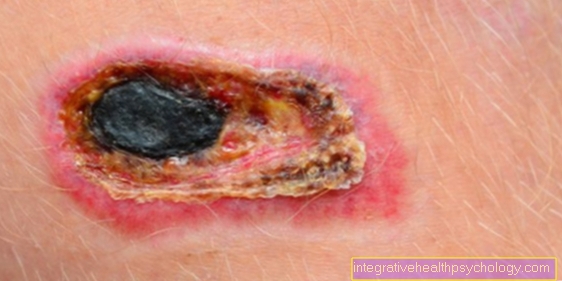
On the one hand, this includes the Accumulation of pus within the gallbladderwhat is known as gallbladder empyema, on the other irreversible destruction of the tissuewhat is known as gangrene.
Ultimately, the Break through the gallbladder wall, a gallbladder perforation occurs.
It can too Accumulation of pus come around. One speaks of a pericholecystic abscess. Liver abscesses are also possible. If the spread is limited, it is a covered perforation. However, the inflammation can also spread systemically as a free perforation in the body and lead to irritation of the peritoneum (peritonitis).
A Spreading to the pancreas as pancreatitis is also possible. Bacteria in the gallbladder can ultimately cause a Blood poisoning (Sepsis) arise. All of these complications are extreme life threatening.
You can also Connecting corridors (Fistulas) to other organs in the abdominal cavity or even in the skin. Gallstones can find their way into the intestines and to the Intestinal obstruction (Ileus) lead. If air from the intestine reaches the bile duct system in the opposite direction, ultrasound detection is carried out using small air bubbles (aerobilia).
Another complication is that crossing acute cholecystitis chronic inflammation of the gallbladder. Chronic gallbladder inflammation can repeatedly cause symptoms through inflammatory flare-ups. In addition, the tissue can change over time in such a way that the Gall bladder shrinks or calcium deposits. The full image is then referred to as a shrink bubble or porcelain gall bubble. Both forms can be in Gallbladder cancer degenerate.
Inflammation of the gallbladder during pregnancy
At about 5% of pregnant women are gallstones to be found and about 1% suffers from complaints.
The formation of gallstones during pregnancy is hormone-related. Especially Estrogens are relevant. In addition, the child growing in the womb can lead to a Displacement of the gallbladder come. The outflow of bile is disturbed and stones can develop more easily.
You might also be interested in: Upper abdominal pain during pregnancy
The composition of gallstones - what are they made of?
The stones are made in one Solution imbalance of bile acids, lecithin and substances such as cholesterol, calcium carbonate, bilirubin.
With about 80% are Cholesterol stones or Mixed stones with a high level of cholesterol the most common Stones. Then follow Bilirubin or pigment stones and Calcium carbonate stonescaused by bacteria, with each 10%.
prophylaxis
Gallstone disease are discovered by chance because they often remain symptom-free for many years. Even with symptom-free gallstone disease, minimally invasive removal of the gallbladder can be used as a prophylaxis for Colic and Inflammation respectively. This variant should be considered, because these stones often cause problems sooner or later. Since the gallbladder is not a vital organ, the body can do without it. The risks of a minimally invasive cholecystectomy are considered to be low.