papilla
definition
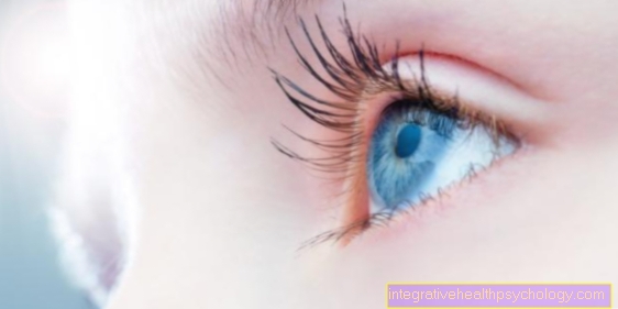
An area on the retina of the eye is called the papilla. All the nerve fibers of the retina converge here and leave the eyeball as a bundled nerve cord in order to be able to pass on the sensory impressions of the eye to the brain.

anatomy
The papilla is a circular area in the retina of the eye and has a diameter of around 1.7 to 2 millimeters, although this can vary from person to person. He's in an ophthalmoscope, also called Ophthalmoscopy referred to as a light, yellowish and circular area to be well delimited from the rest of the retina.
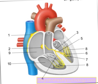
Around one million retinal nerve fibers unite in the papilla and leave the eyeball as a common optic nerve (Optic nerve). After further interconnections, this forwards the visual information of the eye to the brain. In addition, numerous blood vessels enter the eyeball through the papilla and ensure, among other things, the blood supply to the retina.
function
The job of the eye is to convert our visual impressions into information for the brain. To do this, the light falls on the sensory cells of our retina, which then transmits an electrical signal to the nerve fibers connected downstream. These nerve fibers combine in the papilla and emerge from the eye as the optic nerve. This is why the papilla is also known as the optic nerve head.
The papilla itself, on the other hand, has no sensory cells and therefore cannot process visual impressions. It is therefore colloquially known as the “blind spot”. However, as is known, we do not have a black circle in our field of view. The reason for this is that the other eye compensates for this loss and what we see is supplemented into an image in our perception.
Read more about the topic here: Visual field examination
Papillary excavation
The papillae excavation is a cavity in the optic nerve head. An indentation of the papilla occurs, for example, when the intraocular pressure is too high and the nerve fibers that leave the eyeball on the papilla are destroyed due to the long-term excessively high pressure. The cause of this increased intraocular pressure is usually a drainage disorder of the aqueous humor.
The aqueous humor normally has the function of nourishing the lens and cornea. Through its circulation from the rear to the anterior chamber of the eye, it also cleanses the eye of foreign substances and pathogens. For example, if there is a blockage in the so-called Schlemm's Canal, the pressure of the aqueous humor increases on the vitreous humor, which in turn presses on the retina and the papilla. This can lead to the destruction of nerve fibers in the area of the papilla and the areas of the retina from which these fibers originated can no longer transmit information to the brain. As a result, a pathological visual field loss occurs (Scotoma).
The extent of the papillary excavation can be determined by an ophthalmologist by means of an ophthalmoscope, also known as a funduscopy or ophthalmoscopy. Physiologically, there is a certain amount of papillae excavation, which is correspondingly larger in larger papillae than in people with smaller papillae. The ophthalmologist can determine whether it is a pathologically pathological form by measuring the excavation and determining the resulting visual field losses. In addition, the intraocular pressure should be determined, which should be between 10 and 20 mmHg.
Find out all about the topic here: The papillary excavation.

Optic disc edema
The optic disc edema, also called congestion pupil, is a pathological protrusion of the optic nerve head, which is usually slightly arched. In contrast to papilla excavation, the pressure from behind on the optic nerve is increased so that it arches forward.
The causes of papillary edema can be very diverse. In addition to the optic nerve, numerous arteries and veins run through the papilla, which ensure the inflow and outflow of blood for the eye. Therefore, a venous outflow disorder (e.g. central vein thrombosis or sinus thrombosis) can lead to swelling of the optic nerve head.
Another reason can be increased pressure inside the brain skull (intracranial pressure), which can be triggered by masses such as brain tumors, cerebral haemorrhages, infections or inflammations. A congested pupil manifests itself symptomatically as a headache and visual field loss.
To diagnose papilla edema, a reflection of the fundus (funduscopy) should first be performed. If there is a suspicion or finding that is characterized by unclear, blurred boundaries of the papilla as well as a bulging, a comprehensive neurological examination including imaging methods such as computed tomography or magnetic resonance tomography should be carried out to find the cause of the increased pressure.
You may also be interested in: Optic disc edema
Papillary sclerosis
Papillary sclerosis is a hardening of the papillary tissue. In the process, connective tissue in the form of collagen is increased and mostly produced in an uncontrolled manner. The original tissue hardens and loses its elasticity and function. Papillary sclerosis is not an independent disease, but results from another underlying disease. This can be, for example, an inflammation, a circulatory disorder or a degenerative change in the basic tissue.