Spinal ganglia / ganglion cell
Synonyms
Medical: neuron, ganglion cell
Greek: ganglion = knot
Brain, CNS (central nervous system), nerves, nerve fibers
English: nervous system
Explanation
Ganglia are nodular collections of nerve cell bodies outside the central nervous system (= brain and spinal cord). They therefore belong to the peripheral nervous system. A ganglion usually serves as the last switching point before the respective organs into which the nerve processes are to be sent or as the first switching point for nerve processes that are to get from organs to the brain.
It is therefore also an intermediate switching station, where incoming impulses can not only be passed on, but also be "moderated" by other incoming signals. Accordingly, there are motor ganglia for fibers that convey movement information, sensitive ganglia for the transmission of sensory impressions and other sensitive information (pain, tactile, depth sensitivity) and vegetative ganglia in the service of the sympathetic and parasympathetic nerves.
General information can be found at: Ganglion of the nervous system

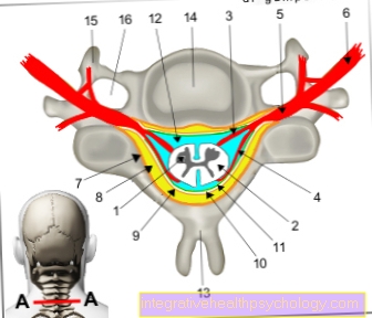
1st + 2nd spinal cord -
Medulla spinalis
- Gray matter of the spinal cord -
Substantia grisea - White spinal cord substance -
Substantia alba - Anterior root - Radix anterior
- Back root - Radix posterior
- Spinal ganglion -
Ganglion sensorium - Spinal nerve - Spinal nerve
- Periosteum - Periosteum
- Epidural space -
Epidural space - Hard skin of the spinal cord -
Spinal dura mater - Subdural gap -
Subdural space - Cobweb skin -
Arachnoid mater spinalis - Cerebral water space -
Subarachnoid space - Spinous process -
Spinous process - Vertebral bodies -
Vertebral foramen - Transverse process -
Costiform process - Transverse process hole -
Foramen transversarium
You can find an overview of all Dr-Gumpert images at: medical illustrations

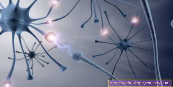
Figure nerve cell
- Dendrites
- Cell body
- Axon
- Cell nucleus
Read more about these topics here: Dendrite and nucleus

Figure nerve cells
- Nerve cell
- Dendrite
A nerve cell has many dendrites, which act as a kind of connecting cable to other nerve cells in order to communicate with them.
tasks
Most ganglia have proper names. Only the segmentally arranged ganglia such as the sensitive dorsal root ganglia, which are located at the level of each vertebra in the intervertebral hole, and the sympathetic ganglia of the trunk are not all named separately.
There are according to the number of processes
- pseudounipolar,
- bipolar and
- multipolar ganglion cells.

Illustration of nerve endings / synapse
- Nerve endings (axon, neurite)
- Messenger substances, e.g. dopamine
- other nerve endings (dendrit)
Both pseudounipolar ganglion cells store the impulses relaying Process (axon, neurite) and the process that brings impulses (Dendrit) directly to each other, so that only a single extension can be seen under the microscope. Pseudounipolar ganglion cells are found in the spinal ganglia, which transmit sensory and sensory stimuli from the body to the spinal cord and brain.
Bipolar ganglion cells have only two cell processes: one Dendrites and a neurite that is often roughly opposite.
Multipolar ganglion cells have in addition to an impulse-transmitting process (axon) at least two, but usually much more impulses receiving processes (dendrites), often hundreds to thousands. They are typical of vegetative ganglia, e.g. in the sympathetic trunk, which is active during stress. Usually all will Ganglion cells of mantle cells (Glial cells) that feed and electrically isolate them.
The spinal ganglia in particular are in close proximity to the Central nervous systembecause they lie in the course of the posterior (sensitive) spinal nerve roots.
They are encased in a capsule-like manner by a protuberance in the membranes of the spinal cord. The nerve cell bodies (somata) and processes of the sensitive nerve cells, but also some blood vessels, are found in the tissue of the spinal ganglion. 80% of the nerve cell bodies are large (around 100 μm) and belong to rapidly conducting "mechanoreceptive" fibers, which are fibers that transmit mechanical effects such as pressure, tension, bending, etc.
The smaller ones (20%) largely belong to the pain fibers.


