Arteries of the neck
introduction

The two major arteries of the neck that supply blood to the head and neck are the subclavian artery and the carotid artery (common carotid artery).
Both give off numerous branches to supply the head and neck organs and the surrounding muscles. They are always set up in pairs: there is one artery for the right side (arteria subclavia dextra or arteria carotis commonis dextra) and one for the left half of the body (arteria subclavia left or arteria common carotis left).
Common carotid artery
The Common carotid artery arises on the right half of the body at the level of the joint between Sternum and Collarbone (Sternoclavicular joint) from a common vascular trunk (Truncus brachiocephalicus) from the artery (aorta), along with the subclavian artery.
On the other hand, they arise on the left half of the body Common carotid artery and the Subclavian artery straight from the bow of the artery (aortic arch).
The Common carotid artery runs together with the Internal jugular vein and the Vagus nerve in the Vascular-nerve street of the necks under the Sternocleidomastoid muscle and next to windpipe as well as the esophagus direction head. In the process, the common carotid artery divides on both sides of the body approximately at the level of the fourth Cervical vertebra into the external carotid artery and the internal carotid artery. This branching is also known as the carotid bifurcation. Both arteries are in their course of one connective tissue membrane enveloped (vagina carotica).
Usually the Common carotid artery do not cut branches before dividing. The glomus organ is found in the area of the carotid bifurcation. The cells of the glomus organ react with increased respiration when the pH value or the oxygen partial pressure of the Blood drops or the partial pressure of carbon dioxide increases.
Internal carotid artery
The beginning part of the Internal carotid artery is widened and contains pressure receptors (baroreceptors) that continuously Blood pressure measure and also detect changes in blood pressure.
The internal carotid artery does not give off any branches in the neck area and runs in the direction of the external carotid artery head. At the Skull base she enters the skull a. The artery and their branches supply the Eye socket, the brain and the Frontal sinus.
In the area of Skull there are important links between Internal carotid artery and External carotid artery as well as the Subclavian artery. These bypass circuits are particularly important when the internal carotid artery slowly closes (e.g. in the case of arteriosclerosis). About the slowly expanding links can supply the Brain With blood then still be maintained. However, a sudden narrowing cannot be compensated for via the collaterals!
There are several important collaterals: One is the Ophthalmic collateral to the external carotid artery, where there is a connection via the Facial artery (Branch of the external carotid artery) and the Opthalmic artery of Eye (Branch of the internal carotid artery). On the other hand, the anastomosis to the external carotid artery and the subclavian artery is over meningeal arteries important. There is a connection from Occipital artery as a branch of the external carotid, the Vertebral artery as a branch of the subclavian artery and meningeal vessels of the internal carotid artery.
External carotid artery
The External carotid artery also pulls direction skull and, with its branches, supplies parts of the Head, of the Facial region and the Meninges. As a rule, it runs in front of the internal carotid artery and crosses the Hypoglossal nerve and the Glossopharyngeal nerve. Overall, there is the external carotid artery in its course 8 branches from.
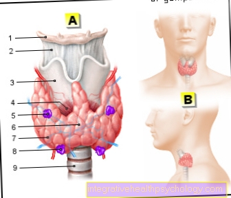
As first branch goes out of the external carotid artery Superior thyroid artery at the level of Hyoid bone (hyoid bone), a bone on the floor of the mouth below the tongue, emerge. She moves to thyroidwhose upper portion they are using blood provided.
Furthermore, through their branch that Superior laryngeal artery, Parts of the Larynx provided.
More branches are closing Muscles above and below the Hyoid bone (infrahyoid and suprahyoid muscles) and to the sternocleidomastoid muscle am neck.
The second branch usually goes above the superior thyroid artery Ascending pharyngeal artery emerges from the external carotid artery. It pulls in the side wall of the Throat (Pharynx), which also supplies them, head towards the Skull base.
Their branches supply them Tympanic cavity of Ear (Arteria tympanica inferior) and the Meninges in the posterior part of the fossa (arteria meningea posterior).
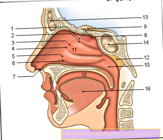
Another branch of the external carotid artery is the Lingual artery. It pulls over the oral cavity to the tonguein which it meanders to the tip of the tongue. In addition to the tongue, it also supplies the floor of the mouth blood.
The arises at the level of the jaw angle Facial artery the external carotid artery. She pulls from Lower jaw in the direction face, at the corner of the mouth and the nose past to the central corner of the eye. It supplies the upper and lower lip of the Mouth.
At the neck gives the facial artery the Ascending palatine artery which supplies blood to the side wall of the pharynx.
Further branches of the facial artery are the Submental artery to supply the Musculature above the hyoid bone and the mandibular salivary gland (submandibular gland) and the Ramus tonsillaristhat draws to the palatine tonsil.
Another branch of the external carotid artery is the Occipital arterythat runs to the back of the head and parts of the lateral and posterior surface of the head as well as parts of the Meninges With blood provided.
The last branch before the external carotid artery divides into its terminal branches is the Posterior auricular artery. She pulls under the Parotid gland in the direction ear and supplies the auricle, middle and inner ear as well as the tympanic cavity of the ear.
At the height of the Jaw angle the external carotid artery divides into theirs Terminal branches, the Maxillary artery and the Superficial temporal artery. The stronger of the two is that Maxillary artery, which gives off a total of 13 branches and the Facial region provided. The weaker end branch is the Superficial temporal artery This draws between the head of the Lower jaw and the external auditory canal in the temporal region. It supplies the parotid gland, parts of the auricle and the external auditory canal. She also gives a branch mimic muscles of the face that Transverse Facial Artery, from.
Subclavian artery
In addition to the common carotid artery, the subclavian artery is one of the large arteries of the neck. In addition to parts of the neck, it primarily supplies the upper extremities and parts of the chest with arterial blood.
As described above, the right subclavian artery arises from the brachiocephalic trunk and the left subclavian artery directly from the aortic arch. It runs over the tip of the lung through a gap between the muscles of the anterior scalenus and the scalenus medius (scalenus gap). This point represents a bottleneck in the course of the artery, at which the blood flow can be impaired. Then it runs on the first rib and under the collarbone towards the shoulder and armpit. There it merges into the axillary artery, which supplies the arm with blood.
In its course, the subclavian artery gives off four branches.
The first branch is the internal thoracic artery that branches off before the scalenus gap and runs along the chest (thorax) to the diaphragm. It supplies parts of the chest wall as well as the pericardium and the diaphragm.
Another branch is the vertebral artery, which runs to the sixth cervical vertebra and runs between the cervical vertebrae with the vertebral vein and the vertebral nerve up to the head. It enters the head in the posterior fossa of the skull and unites with the vertebral artery on the opposite side to form the basilar artery. This supplies parts of the brain and spinal cord.
The next branch is the thyrocervical trunk, a trunk of the subclavian artery from which three arteries arise: the inferior thyroid artery, the suprascapular artery and the transversa cervical artery (or transversa colli artery).
The inferior thyroid artery crosses the vascular-nerve road of the neck and runs to the back of the thyroid gland, which supplies it with blood. The inferior laryngeal artery, which runs to the larynx, and the ascending cervical artery, which run together with the phrenic nerve towards the head and supply parts of the neck muscles and the spinal cord, emerge from it.
The suprascapular artery runs behind the collarbone (clavicle) to the shoulder blade and supplies not only the collarbone and shoulder joint but also some surrounding muscles. At the lateral edge of the shoulder blade, it connects (anastomosis) with branches of the axillary artery.
The arteria transversa cervicis has branches that supply the neck, neck and shoulder muscles. In addition to the thyrocervical trunk, the costocervical trunk also emerges from the subclavian artery. Two branches arise from it: the deep cervical artery, which runs towards the head and, in addition to the deep neck muscles with branches (rami spinales), also supplies the membranes of the spinal cord, as well as the suprema intercostal artery for supplying the upper muscles in the spaces between the ribs (intercostal muscles).