Bile duct cancer
Synonyms in the broadest sense
Bile duct carcinoma, biliary tract tumor, biliary tract carcinoma, cholangiocellular carcinoma (CCC), cholangiocarcinoma, biliary cancer, Klatskin tumor, hilar cholangiocarcinoma
Note
All information given here is only of a general nature, tumor therapy always belongs in the hands of an experienced oncologist (tumor specialist)!
definition
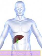
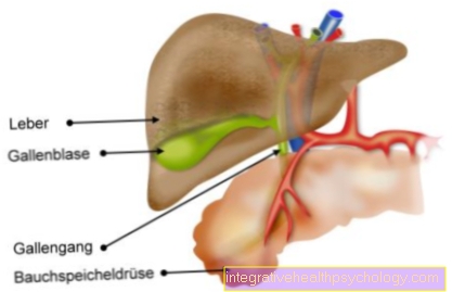
The bile duct tumor is caused by the degeneration of the bile duct mucosa into an uncontrollably growing, malignant tissue (carcinoma). Bile duct cancer (bile duct cancer) grows relatively slowly and spreads (metastasizes) into other tissues relatively late. In bile duct cancer, a distinction is made between tumors that arise in bile ducts that are inside (intrahepatic) or outside (extrahepatic) the liver. Overall, bile duct cancer has a poor prognosis, i.e. it is often no longer curable at the time of its diagnosis. A special form of bile duct carcinoma is the Klatskin tumor, which arises at the confluence of the excretory ducts of the right and left lobes of the liver in the common hepatic duct (common hepatic duct).
frequency
Bile duct carcinomas are very rare overall. Gallbladder cancer is 3 to 5 times as common as bile duct cancer. The peak of the disease is beyond the age of 60. Men are more often affected by tumors of the biliary tract, in contrast to gallbladder cancer, which is more likely to affect women.
Tumor types and location
In the case of bile duct cancer, it is mostly (histologically) fine tissue Adenocarcinomas, that means that the tumors originate from gland cells in the bile ducts. The tumor develops in a ring around the duct and then longitudinally along the bile ducts. In the further process of the disease, the cavity (lumen) of the duct narrows and the bile builds up in the duct liver. As a result, a Jaundice (Jaundice) developed. The tumors tend to develop Forks the biliary tract, for example at the confluence of the excretory ducts of the left and right lobes of the liver into the large common duct (ductus hepaticus communis). Bile duct tumors that arise in this area will be Klatskin tumors called. Another site of predilection for tumor development is the confluence of the common hepatic duct with the duct of the Gallbladder (Ductus cysticus).
Causes and Risk Factors

The development of gallbladder cancer is favored by various risk factors.
Autoimmune diseases like Ulcerative Colitia, an inflammatory bowel disease and primary sclerosing cholangitis (PSC), a chronic inflammatory biliary tract disease caused by the proliferation of connective tissue with constrictions (Strictures) associated with the biliary tract correlate with the occurrence of biliary tract tumors. Patients with these diseases have a thirty-fold increased risk of developing biliary tract cancer.
Another predilection factor is innate Caroli syndromewhich is associated with sack-like bulges in the biliary tract that lie within the liver (intrahepatic common bile duct cysts).
Also through infections of the biliary tract with parasites like Liver fluke and Trematodes the development of this type of cancer is favored.
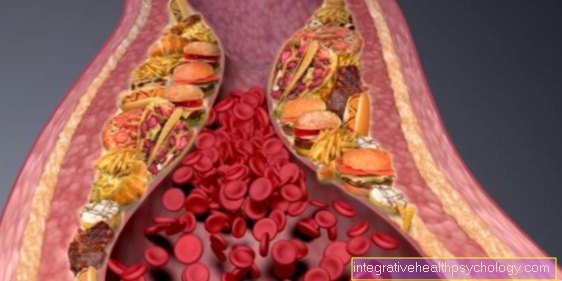
In addition, there is a connection with the chronic consumption of Cigarette smoke observed. In particular, that which occurs in cigarette smoke is intended here Dimethylnitrosamide which play an important role as a carcinogenic substance.
In contrast to gallbladder carcinoma, correlate Gallstones not with the appearance of the bile duct tumors.
Symptoms
The cardinal symptom is one painless jaundice (Jaundice), which is caused by the narrowing of the bile ducts, causing the bile to build up in the liver. The accompanying symptoms of jaundice are one Yellowing of the skin and the white Eye color (Dermis, sclera) and an onerous itchy skin as a result of bile salts deposited in the skin. There is also a clay-like Discoloration of the stool by the lack of bile pigment in the stool and one Urine dark in colorbecause the kidney takes over the excretion of the bile pigments. Due to the lack of bile acids in the small intestine, fats can be digested more poorly, resulting in intolerance to high-fat meals and too Fatty stools (Steatorrhea) can come.
When the tumor closes the duct of the gallbladder (ductus cysticus), the bile is retained in the gallbladder. In addition to the painless jaundice, a bulging gallbladder can be felt under the right costal arch. This symptom complex is also called Courvoisier's symbol designated.
More ailments can turn out to be unspecific diffuse Upper abdominal pain, nausea, Vomit, Loss of appetite and Indigestion represent. As a late sign can Pain in the upper right abdomen and other non-specific symptoms that can occur in many cancers, such as Weight loss (Tumor cachexia), Anemia (anemia), Tiredness and listlessness.
The congestion in the biliary tract can easily become dangerous Infection of the biliary tract (Cholangitis), as the “standing” bile is a suitable breeding ground for bacteria.
In the course of the accumulation of the bile it can become one Liver failure (Hepatic insufficiency) and in the final stage to the complete loss of the Liver function With coma and severe bleeding disorders come.
Tumor spread (metastasis)
Different forms of metastasis are described here:
- Lymphogenic metastasis:
The lymph vessels drain the lymph fluid from all parts of our body. If the tumor is connected to a lymphatic vessel as it grows, it can easily happen that some cells become detached from the tumor cell cluster and are carried away with the lymph flow. There are numerous lymph nodes in the course of a lymphatic vessel. In them is the seat of Immune defensewhich has the task of catching and fighting germs (bacteria). The tumor cells settle in the nearest lymph nodes and multiply again there. This is how a Lymph node metastasis. In this type of cancer, lymph nodes that are in the immediate vicinity, i.e. in the vascular compartment of the liver (Hepatic hilum), and later also in those in the course of the main artery (aorta). This type of cancer often already shows lymph node metastases at the time of diagnosis, so it is always advisable to remove the surrounding lymph nodes during the operation.
- Haematogenic metastasis:
If the bile duct cancer gets attached to a blood vessel as it grows, cells can tear themselves loose in this situation and be spread throughout the body via the bloodstream. As the first station, the blood flows through the liverwhere the carcinoma cells can settle and Daughter tumors (Distant metastases) can form. In the further course of the disease, cells can also detach themselves from the liver metastases and continue into the lung sprinkle. Later metastasis can also develop in the Peritoneum (Peritoneum), this is then also peritoneal carcinosis (Peritoneal cancer) called.
- Per continuitatem:
The bile duct cancer can grow into other neighboring organs as it spreads (tumorous infiltration). Depending on the proximity of the carcinoma to the liver, the tumor may have grown into the liver when the diagnosis was made. In addition, the tumor can in the further course of the Duodenum (Duodenum), stomach, pancreas (Pancreas), adjacent vessels such as the portal vein (Vena portae) and other adjacent structures grow into it.