hip joint
General
Humans have two hip joints that are symmetrically arranged and are responsible for leg movements as well as for dissipating the forces acting on the body. Furthermore, the hip joints, together with the spine, take on the main tasks of the body's statics. Numerous ligaments secure the actual hip joint, and the muscles anchored to the thigh also provide additional security and stability.

Structure of the hip joint
Like any joint, the hip joint also has one Swivel head and a Socket. Roughly one can say that the socket in the Pelvic bones represents a kind of hemispherical recess. The joint head is through the Femoral head formed, which dips into the joint socket.
According to the definition, the hip joint is called the Facies lunata acetabuli at the Hip bone formed, as well as from caput femoris (Femoral head). The face is the lining of the described hollow sphere on the hip bone. In order to ensure stability, the joint head must be securely held in the socket.
In the case of the hip joint, the Femoral head is larger than the joint socket. For this reason, the joint socket is anatomically enlarged by an enlargement and thus ensures a more secure fit of the Thigh in the pan. The magnification is also called Labrum acetabuli or also called joint lip. The joint lip also exists Fiber cartilage. Together with the facies they cover 2/3 of the joint head and thus ensure its stability.
As Pan roof one understands the middle part of the upper edge of the pan. It is condensed and can be represented well in the X-ray image. This pulls on the lower part of the pan Transverse acetabular ligament, which also contributes to the stability of the hip joint. The Acetabular fossa is designed with a body of fat, which should ensure a lower-friction movement and have a cushioning effect in the event of impacts
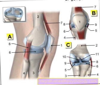
Illustration of a hip joint

- Acetabulum - Acetabulum
- Iliac scoop - Ala ossis ilii
- Iliac crest - Iliac crest
- Articular lip of the acetabulum -
Labrum acetabuli - Femoral head (= femoral head) -
Head femoris - Femoral neck - Collum femoris
- Great Rolling Hill -
Greater trochanter - Thigh shaft -
Corpus femoris - Small rolling hill -
Lesser trochanter - Ischium - Os ischii
- Hip hole - Obturate foramen
- Pubic bone - Pubis
- Lumbar and sacrum kink -
Promontory
Three bones fuse to form the hip bone:
Ilium, pubic bone and ischium
You can find an overview of all Dr-Gumpert images at: medical illustrations
Appointment with a hip expert?

I would be happy to advise you!
Who am I?
My name is I am a specialist in orthopedics and the founder of .
Various television programs and print media report regularly about my work. On HR television you can see me live every 6 weeks on "Hallo Hessen".
But now enough is indicated ;-)
The hip joint is one of the joints that are exposed to the greatest stress.
The treatment of the hip (e.g. hip arthrosis, hip impingement, etc.) therefore requires a lot of experience.
I focus on treating all hip diseases in a conservative way.
The aim of all treatment is treatment without surgery.
Which therapy achieves the best results in the long term can only be determined after looking at all of the information (Examination, X-ray, ultrasound, MRI, etc.) be assessed.
You can find me in:
- - your orthopedic surgeon
14
Directly to the online appointment arrangement
Unfortunately, it is currently only possible to make an appointment with private health insurers. I hope for your understanding!
You can find more information about me at
bone
The hip joint consists of the femoral head (joint head) and the hip joint bone (joint socket). The so-called caput femoris is the ball that delimits the upper thigh bone. The thigh neck closes at it (Collum femoris), which then represents the transition to the actual femur.
The femoral neck is often affected by fractures, especially in the elderly.
The pelvis is the largest bone in the human body. It is very massive and, together with the spine, supports the human being. The pelvis consists of three sections that are fuzzy from each other and the hip bone (Os coxae) represent. These parts are called the pubic bone (Pubis), Iliac bone (Os ilium) and ischium (Os ischii) designated. In the area where the three anatomical sections come together, you will find the acetabular fossa, the socket for the hip joint. The fossa is delimited by the Facies lunata, which gets its name from its crescent-shaped appearance.
There is also a small bony indentation in this area (Acetabular notch). The acetabular limbus wraps itself in a circle around the joint socket and delimits it to the outside.
Tapes
The hip joint is secured by numerous ligaments. The strongest Band of the human body represents that Iliofemoral ligament It has a load capacity of 350 kg and has its starting point at the hip bone and then pulls itself slightly turning outwards down to the thigh bone, where it has its second starting point on the upper part.
Overall there is five Ligaments at the hip joint. Four of them are outside the joint and one is inside. The ligaments on the outside make it up Ring band, which is also called Zona orbicularis referred to as.
The following ligaments belong to the section located in the joint: the Ischio-femoral ligament pulls from the os ischi to the head of the femur, the Pubofemoral ligament from the pubis and that Iliofemoral ligament from the ileum to the head of the femur. The ligaments of the hip joint have two main roles. On the one hand they stabilize and strengthen the joint, on the other hand they limit the range of motion and prevent unphysiological movements in the hip joint. The Ring band loops around the narrowest part of the hip joint and acts as a very strong one stabilizer. The head of the thigh is in the ring ligament and is held by it.
This is the only ligament that runs in the joint Ligamentum capitis femoris. The areas that are not secured by tapes are considered to be endangered because the stability there is severely limited and Fractures or a "Dislocate“Of the joint can take place there.
Capsule:
A joint capsule is a tough skin that surrounds each joint and rests closely on the joint and protects it or substantially to the joint Joint stability contributes. In the hip joint, the joint capsule is attached to the outside of the acetabular labrum and to the hip bone. The acetabular labrum protrudes freely into the capsule.
capsule and Cartilage margin run approximately at the same level, the area of the femoral head neck that is not enclosed by the joint capsule is shorter at the front than at the back. The attachment lines of the joint capsules run close to the anatomical structures of the hip joint. The so-called Linea intertrochanterica should be mentioned in the front area and the Crista intertrochanterica behind, more precisely, the capsule attachment line is about 1 cm away from it.
Vessels
Like any bone, the bones of the hip joint are covered with blood leading to the bones Blood vessels, provided. In the area of the head of the femur, vessels enter the femur on each side, which are called Arteriae capitis femoris are designated.
Tears or clamps can mean harmful undersupply of the bone and must be excluded with every injury and every fracture. In addition to supplying the Thigh the artery also supplies those passing in this area Tapes. The pelvis is made up of the smallest arteries, followed by the large ones Arteries branch off, supplied.
Muscles
The numerous muscles, which, in addition to stabilization, also take on the task of moving, make a significant contribution to the stability of the hip joint. A distinction is made between the flexors, extensors, abductors and adductors of the hip muscles.
- Straightener: to the stretchers count the Glutes (Gluteus maximus muscle, M. gluteus minimus and M. gluteus minimus), the M. adductor magnus and the piriformis muscle. In addition to stretching the hips, they are also responsible for stability in the same.
- Flexor: at the inflection they are Muscles iliopsoas, tensor fasciae latea, pectineus, adductor longus, brevis and Gracilis muscle involved.
- Abductors: for the abduction, i.e. the splaying of the thigh, the muscles gluteus medius, tensor fasciae latea, gluteus maximus, minimus, piriformis and obturatorius are responsible.
- Adductors: the reattachment of the leg (adduction) is done by the muscles adductor magnus, longus, brevis, Gluteus maximus muscle, gracilis, pectineus, M. quadratus femoris, and obturatorius externus.
Together, the muscles push the head of the femur into the acetabulum and thus contribute to the stability and resilience of the hip joint.
annoy
Numerous nerves also move around the hip joint and mainly serve the sensitive care the hip muscles. Parts of the muscles are derived from the direct nerve branches Spine supplied (L1-L3 and L2-L4). Furthermore, the Superior gluteus nerve, Inferior gluteus nerve, Sacral plexus and the Obturator nerve in the area of the hip.
As with the vessels, injuries and fractures must always be checked to see whether a nerve has been injured. Typical symptoms of paralysis of muscles supplied by the corresponding nerves indicate the location of the damage.
The different angles of the hip joint
In the hip joint they can External rotation, Internal rotation, the diffraction, the Elongation, the Spreading (Abduction) and the Reinstall (Adduction) be performed. There are also numerous mixed movements that are possible in the hip joint. The femoral head is at a certain angle in the acetabulum. This angle depends on the Age and changes with age.
In the 3-year-old child the angle is 145 degrees, in the adult it takes up 126 Degrees, and in the elderly the angle is only 120 degrees. The reason for this is the different stabilities and stages of ossification at the corresponding age.
Furthermore, there are numerous diseases and malpositions in which the angle also changes. With the known Bow legs (coxa vara) can be the angle 90 Degrees, while with the Knock knees (Coxa valga) the angle almost 160-170 Degrees. Basically, angles between 120 and 145 degrees are the most stable. Since the angle changes progress slowly and not suddenly, the body compensates for this instability through active bone remodeling and additions. The different angles not only affect the stability in the hip joint, but also have a minor effect on the ability to move.
This is how people with an angle (also known as Collum-corpus angle) of 126 degrees perform the full range of possible combinations of movements in the hips, while very old people with an angle of 120 degrees are restricted in a large number of movements possible in the hips for mechanical reasons. It is not clear whether a decrease in the collum-corpus angle can also lead to a higher susceptibility to fractures.
Summary
The hip joint is that greatest Joint of the body that is shared with the Spine makes a significant contribution to the stability and statics of the body. The hip joint, also known as the articulatio coxae, is made up of the Femoral head, which represents the joint head and the Hip boneshowing the socket with a crescent-shaped notch. To ensure sufficient stability in the joint, it is important that the joint head fits exactly into the joint socket. In the case of the hip joint, the femoral head is larger in relation to the socket. In order to guarantee the stability nevertheless there is an anatomical one Socket enlargement, which are also called Joint lip referred to as.
The hip joint is made up of numerous Tapes and Muscles stabilized. The ligaments that stabilize the hip joint run from the hip bone to the thigh. The most important ligaments in this area are the ileofemorale ligament, the ischiofemorale ligament, and the pubofemorale ligament. Together they form the so-called Ring bandthat holds the femoral head like a button in a buttonhole. One of the 5 hip ligaments runs within the joint and is also known as the ligamentum capitis femoris. Its joint capsule, which also has a stabilizing effect, surrounds the femoral head and joint socket.
Numerous Muscles in and around the hip joint ensure that all possible movements can be carried out and also act as stabilizers in the joint. The most important muscles, along with some others, are the Gluteus maximus muscle, medius and minimus.
In addition to the small ones Arteriesthrough which the hip joint is supplied with blood, there is an artery flowing into the head of the femur, also known as the arteria capitis femoris. In the event of injuries or accidents, it is always important to check whether the vessels have been injured. In this case a not inconsiderable bleeding on the one hand but also massive undersupply of the hip and thigh bones on the other hand must be feared. The same applies to injuries to the nerves supplying the hip muscles, which must also be checked for integrity after an accident.
The head of the femur is at a very specific angle in the hip joint. This angle depends on, among other factors, the Age. Newborns and young people have an angle of approximately 145 degrees, adults have an angle of approximately 126 degrees, and for old people the angle is approximately 120 degrees. The older a person gets, the steeper the head of the femur stands in the hip joint. There are still some diseases in which the angle is also changed. At Bow legs (Coxa varum) the angle is closer to 90 degrees, while with the Knock knees (Coxa-valga) the angle becomes steeper and can be around 170 degrees. Hip joints, the angles of which are either very steep or very flattened, have a certain angle compared to the normal angles instability on. Due to the slow development, the body can initially compensate for the instability.