Meninges
Synonyms
Medical: Meninx encephali
English: corneal
definition
The meninges are a layer of connective tissue that surrounds the brain. In the spinal canal, it passes into the skin of the spinal cord. Humans have three meninges.
From the outside in, these are the hard meninges (Dura mater or Leptomeninx encephali), and the soft meninges (Pia mater or Pachymeninx encephali), as well as the cobweb skin (Arachnoid mater), which lies in between.

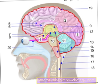
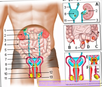
Illustration of the meninges

Meninges
- Tendon hood -
Galea aponeurotica - Emissary vein -
Vena emissaria - Hard meninges (dura) -
Cranial dura mater - Cobweb skin of the brain -
Arachnoid mater cranialis - Soft meninges (pia) -
Pia mater cranialis - Gray matter -
Substantia grisea - White brain matter -
Substantia alba - Diploevene - Diploic vein
- Skull roof - Calvaria
- Villi of the cobweb skin -
Granulationes arachnoideae - Subdural gap -
Subdural space - Cerebral sickle - Falx celebri
A - roof of the skull and meninges
schematic frontal section
You can find an overview of all images from Dr-Gumpert under: medical images
function
There are three different meninges that surround the brain and perform different tasks.
In general, the meninges are used to protect the brain. The spaces between them absorb shocks and changes in volume. They also play an important role in supplying nutrients to the nerve cells of the brain.
The hard meninges on the outside (Dura mater) mainly used to protect the brain. It also contains blood vessels in its invaginations that drain blood from the brain. There are many pain receptors in the hard meninges, which is why they are very sensitive to pain.
The so-called spider web skin (Arachnoid) contains many smaller blood vessels that supply the brain. In addition, it fulfills the function of the exchange between the cerebral fluid (Liquor) and the blood. Here, the cerebral fluid is absorbed in the area of special protrusions of the meninges (arachnoid villi) and passed on to the draining blood vessels.
The soft meninges are closest to the brain tissue (Pia mater). It is used to supply the brain tissue with nutrients.
Also read the article on the topic: Choroid plexus
Leptomenix - dura mater

anatomy
The Dura mater forms a rough skin between the Skull bones and the Brain surface. It divides into two leaves, the outer leaf at the same time being the inner periosteum (Periosteum) of Skull forms and the inner sheet with the Cobweb skin (Arachnoid) merges. So between the hard meninges and the skull bone exists under physiological conditions no space in between. A so-called Epidural space however, can be under pathological conditions form, such as through Bleeding or Dream. There is one in the spinal cord area physiological epidural spacewhich is filled with adipose tissue.
The hard meninges does not nestle in the individual indentations and coils (Gyri and Sulci) of Brain, but it forms the so-called at larger gaps Dural septa out. The largest septum is the Falx cerebri, which runs crescent-shaped in the middle of the upper skull from front to back and the two Cerebral hemispheres separates. Both of them too Cerebellar hemispheres (Cerebellum) are separated by a dural septum, the Falx cerebelli is located in the back of the skullcap.
Below the Pituitary gland the hard meninges form that Sellae diaphragm with an opening for the style of the pituitary gland. Between the Occipital lobe (occipital lobe) of the cerebrum and the cerebellum, it finally forms the tent-shaped one Tentorium cerebelli.
In addition to the dura septa, the hard meninges form so-called sinuses through duplicates, which have a surface lining similar to that of blood vessels. They act as venous blood collection vessels that carry the blood from the meninges and the brain to the Internal jugular vein derive. The most important are the Superior sagittal sinus in the upper margin, and the Inferior sagittal sinus at the bottom of the Falx cerebri and the Transverse sinus, which runs semicircular in the rear, lower skull base area.
function
The hard meninges serves to protect the brain tissue by mechanically stabilizing it during rapid movements or trauma. Furthermore, it contains large, efferent blood vesselsthat controls the outflow of blood from the brain via the Jugular vein in the Superior vena cava and so ins heart guarantee.
Pachymeninx - arachnoid

anatomy
The Cobweb skin forms a fine layer under the Dura mater, on the underside of which it hugs completely. So it also shapes everyone Dural septa With. So there is no subdural space per se. However run below the translucent Arachnoid the blood vessels on the surface of the brain. The fine veins that that blood from the brain transport away, step on a short distance through the Arachnoid and the inner sheet of the dura materto get into the Sagittal sinus and the Transverse sinus to get. These vessels that Bridging veins, may tear and bleed, causing a Subdural bleeding (Cerebral hemorrhage) and creates a gap between the dura mater and the cobweb skin. Below the Cobweb skin lies the physiological subarachnoid spacewho the outer cerebrospinal fluid space represents. So this is where it flows Nerve water (Cerebrospinal fluid), which cushions the brain and also the spinal cord during jerky movements or jolts. The Subarachnoid space is divided by connective tissue septa that connect the arachnoid with the underlying pia mater. Between these septem the superficial blood vessels of the brain run in the subarachnoid space.
function
The Arachnoid Fulfills two important tasksthat are essential for the proper functioning of our brain. For one thing, it educates fine protuberanceswhich extend through the inner sheet of the meninges into the sinus veins. These so-called Pacchioni granulations (Granulationes arachnoideae) absorb the CSF from the subarachnoid space and release it into the sinus veins in the dura mater. Through the Choroid plexus in the inner liquor space always new nerve water formed so that the liquor is constantly circulated and renewed. Furthermore, the upper layer, which is in direct contact with the dura, forms the Blood-brain barrier.
By tight junction, i.e. very closely fused cell connections, a barrier is created through which no blood components can pass into the nerve water. This is so important because some substances in the blood are used for the Toxic nerve tissue would be (poisonous). Many too Medication cannot pass the blood-liquor-barrier and have to be modified extra molecularly in order to be able to work in the brain.
Pia mater
anatomy
The pia mater forms the innermost layer of the meninges. It lies directly on the brain tissue and also follows its coils and crevices. It forms a layer of connective tissue around the blood vessels entering the nerve tissue and accompanies it into the interior of the brain.
Innervation and blood supply to the meninges
The blood supply to the meninges occurs through the Anterior meningeal arterythat supplies the front part that Meningeal artery mediathat supplies the middle part, as well as through the Posterior meningeal arterywho is responsible for the rear portion.
All three arteries are branches of the external carotid artery (External carotid artery).
The brain itself is made up of branches of the internal carotid artery (Internal carotid artery) provided.
The innervation of the meninges occurs predominantly via the Trigeminal nerve, the fifth cranial nervewho is also for the sensitivity (Sensitivity to pain and pressure) of the face is responsible. A small rear portion is dated Vagus nerve provided.
In contrast to the brain, everyone is three meninges extremely sensitive to pain.
Pain
The Meninges play a crucial role in Pain development in the head. Since that Brain itself no pain receptors it is insensitive to pain. The meninges, on the other hand, have many such receptors and are therefore particularly sensitive to external pain stimuli.
Often pain is caused by too strong pressure on the meningeslike he did with a Bleeding of the brain occurs.
But also one Inflammation of the meninges (meningitis) can lead to severe pain.
Tension
Tension in the upper back and / or neck area are a common cause of a headache. Also tension of the Masticatory muscles can lead to this.
In extreme cases, you can through this Tension in blood vessels or nerves trapped become. Is it due to it Circulatory disorders of the brain or the Meningesso the pain can be very intense.
Long-lasting or very strong tension should therefore, especially if they cause strong symptoms, from one Physiotherapist or an osteopath be treated.
Meningitis
The Meningitis is in the jargon meningitis called. It is an inflammation caused by various pathogens, such as Viruses, bacteria or fungi, can be triggered. These can get to the meninges through the blood and especially in people with a weakened immune system cause infection.
In most cases, meningitis is caused by Viruses caused. Bacterial meningitis is not that common, but it is much more dangerous. It can be life-threatening within a few hours. It is usually triggered by Meningococci or Pneumococci.
The so-called TBE is a special form of meningitis in which also affects the brain is. This is through Ticks triggered.
At the beginning, meningitis often manifests itself as sudden onset Flu symptoms. Those affected complain about Headache, fever, body aches and chills. However, come Neck stiffness and Pain in the neck when moving the head towards the chest added. This is because with this movement the meninges become tense and this leads to severe pain.
Also one Photophobia can occur.
Should these symptoms occur a doctor should be consulted immediately. Occur in children often small bleeding in the skin (Petechiae) added.
Often times, meningitis in children is associated with a Viral disease, how mumps, chickenpox or measles, to observe.
Because meningitis life threatening can be, it is very important to children vaccinate adequately allow. Especially since the disease often has a severe course, especially in infants and children. This is mainly due to the not fully trained immune system traced back.
It is very important that the doctor quickly finds out which trigger is causing it Meningitis has come. Only then can appropriate therapy be started promptly. Blood draw and a Lumbar puncture (Removal of Nerve water) are important steps in diagnosis. So it can be found out if it is bacterial or viral pathogens acts.
A Computed Tomography (CT) or one Magnetic resonance imaging (MRI) can provide information about the current State of the brain deliver.
In the case of meningitis, the patient is admitted to the hospital as an inpatient. Depending on the pathogen, it will with Antibiotics (in the case of bacteria) treated or merely symptom-oriented if it is a viral meningitis. If there is a bacterial infection, all contact persons of the patient are treated with antibiotics as a precaution.
If therapy is started in time, meningitis usually heals without consequences. However, in some cases it can too neurological sequelae come. These can be found in Hearing damage, Signs of paralysis or even in Behavior changes express.
Often there are consequential damages, if the meningitis has already spread to the brain.
Meningeal irritation
The meninges are innervated by sensitive nerves and are therefore sensitive to pain. For this reason, irritation of the meninges can trigger symptoms such as headaches.
Causes of meningeal irritation
Meningeal irritation can have different causes: For example, a Sunstroke cause irritation of the meninges. Often, however viral infections Cause of such irritation. Also Bacteria or fungi can lead to the typical symptoms.
Meningeal irritation symptoms

Complaints can include headaches, painful stiff neck, nausea, sensitivity to light and noise. If the neck stiffness is due to irritation of the meninges, it is called Meningism. Neck stiffness is caused by reflex and arises from the fact that when the head is bent forward, the meninges are stretched and this triggers head and neck pain in the person concerned. Depending on the degree of irritation, this pain can already be present at rest. The symptoms are similar to those of meningitis (meningitis) and therefore require medical clarification.
Read more on the subject at: Meningeal irritation
In the case of neck stiffness that is not based on irritation of the meninges, e.g. in the case of diseases of the cervical spine, another therapy is required.
An important differential diagnosis, a disease that causes symptoms similar to meningeal irritation, is migraine.
Diagnosis of meningeal irritation
Certain clinical signs and laboratory values can provide information and provide clues about the underlying disease. Atypical symptoms can appear in children, such as resting the hands to the side when sitting, knees and hips remain bent, the so-called. Amoss mark. The doctor will check whether there is pain or stiffness in the neck when bending the head (menigism sign). In children, the knee kiss and tripod can be noticeable, in adults it can Brudzinski, Kernig and the Lasegue symbols be.
An examination of the brain water can also provide information about a possible disease. To do this, a Lumbar puncture carried out.
Therapy of meningeal irritation
The duration and treatment of the meningeal irritation depends on the root cause or the underlying disease. Come with bacterial inflammation Antibiotics used, for viral pathogens, antiviral drugs. Also masses in the area of the meninges, e.g. metastases, can cause stretching and thus also lead to irritation of the meninges. A distance the mass is then necessary to alleviate the discomfort.
In general, however, there will be above all one Pain management started to relieve the headache that occurs.
Meningeal injury
Depending on which area of the meninges is injured, follow different consequences and different treatments are required:
Between the Cobweb skin, so the so-called arachnoid mater, and the hard meninges, so-called dura mater Bridging veins. If there is an injury in the area of these veins, venous bleeding occurs, too Subdural bleeding called. Since there is only a low pressure in the veins, bleeding from the bridge veins is also significant slower as a bleeding from arteries. Symptoms such as visual disturbances, dizziness and severe headaches only appear after a few hours.
If, on the other hand, an injury occurs under the spider web skin, one speaks of one Subarachnoid hemorrhage (sub= Latin for under). Since there are mainly arteries in which there is a high pressure, symptoms appear here within seconds and it is an urgent one medical emergency.
Are those arteries affected that supply the meninges themselves, e.g. the A. meningea media, it is also an arterial hemorrhage. Since these arteries run between the hard meninges and the skull bone, a space is created here which, under normal circumstances, does not occur in the skull. This is called bleeding "Epidural bleeding"This bleeding should also be treated medically as soon as possible.
Metastases in the meninges
In the case of tumor diseases, cells can “wander” via the blood and lymph vessels and settle in another part of the body. This process is called metastasis and the resulting tumor deposits are accordingly metastases.
Meninges metastases can grow in the skull and trigger symptoms similar to those of brain tumors. Since different areas in the brain perform different tasks, the metastases of the meninges occur depending on the exact location and size of the metastases different failures. The treatment is similar to that of a brain tumor and the prognosis depends on various factors, including the time at which the main tumor spread, known in technical terms as the primary tumor. If this has spread late, there is a better prognosis. Most of those affected die from the primary tumor and not from the brain metastases.


.jpg)