Impingement syndrome of the shoulder from a physiotherapeutic point of view
Note
The medical-orthopedic part can be found under our topic Impingement Syndrome, written by .
Synonyms
- Shoulder constriction syndrome
- Tight shoulder
- painful shoulder
- painful arch
- subacromial impingement
- subacromial tightness
- PHS = humero scapular periarthritis

definition
The term Impingement Syndrome Derives from the Anglo-American usage and means something like bump into, entrapment, with the shoulder usually meaning an entrapment between the ball of the humerus head and the bony shoulder roof.
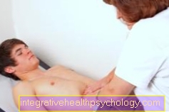
Appointment with a shoulder specialist

I would be happy to advise you!
Who am I?
My name is Carmen Heinz. I am a specialist in orthopedics and trauma surgery in the specialist team of .
The shoulder joint is one of the most complicated joints in the human body.
The treatment of the shoulder (rotator cuff, impingement syndrome, calcified shoulder (tendinosis calcarea, biceps tendon, etc.) therefore requires a lot of experience.
I treat a wide variety of shoulder diseases in a conservative way.
The aim of any therapy is treatment with full recovery without surgery.
Which therapy achieves the best results in the long term can only be determined after looking at all of the information (Examination, X-ray, ultrasound, MRI, etc.) be assessed.
You can find me in:
- - your orthopedic surgeon
14
Directly to the online appointment arrangement
Unfortunately, it is currently only possible to make an appointment with private health insurers. I hope for your understanding!
You can find more information about myself at Carmen Heinz.
Symptoms
In the range of approx.60-120 ° splaying or lifting movement of the upper extremity, sharp pulling pains usually occur in the front, lateral area of the upper arm, which are so severe that they result in a change in the sequence of movements or an interruption in movement.
If the movement is continued, the pain subsides or disappears completely, as the position of the shoulder joint head relative to the shoulder roof changes (it increasingly goes into external rotation). This phenomenon is known in the technical language as "painful arc".
The pain is caused by the pinching of connective tissue structures (tendons, capsule parts, bursa) in the anatomically narrow tunnel between the shoulder joint head and the shoulder roof (acromion). This tunnel narrows when the arm spreads out by approx. 60 °. Cleaning windows, blow-drying your hair, putting things in the closet or grabbing the seat belt can be a pain. The pain is mostly localized in the area of the lateral upper arm, whereby the deltoid muscle located there is not causally affected, sometimes it radiates towards the elbow or shoulder blade. If it radiates up to the elbow joint, it is likely that the cervical spine is responsible as a pain intensifier.
Usually the pain is stronger with active movements, which are carried out by the person concerned, than when the shoulder is passive e.g. is moved by a doctor or therapist. Furthermore, the quality of the pain depends on the way in which the movement is carried out. A small change in shoulder position in the direction of inward or outward rotation can increase or decrease the pain during the spreading movement.
Often there is also pain at night, the patient can no longer lie pain-free on the affected shoulder.
Read more on the topic: Inflammation of the shoulder blade
causes
The cause of the pinching of tendons under the roof of the shoulder (to impinge = pinch, bump) can be either in the connective tissue structures of the shoulder joint (tendons, capsular ligamentous apparatus) or in the bony parts.
Connective tissue causes
The main cause is the tendon of the supraspinatus muscle, which extends up to 1 cm through degenerative processes. can swell, and is then disturbed in their running and sliding behavior. However, other tendons of the shoulder muscles (predominantly the tendons of the so-called Rotator cuff (Torsional muscles of the shoulder joint, which center the shoulder head in the joint when tensioned), ligament connections, portions of the shoulder joint capsule or the bursa under the bony shoulder roof.
Another cause is the so-called Shoulder joint instability. This means that the joint is not able to assume an anatomically correct position between the head and the socket during normal movements and loads. In 85% of the cases the shoulder joint head slipped upwards and forwards (subluxation, is favored by the anatomy) and then leads to the tendons being pinched when the arm is raised.
Even a minimal shoulder instability results in increased activity of the shoulder joint muscles, which leads to tension, trigger points and ultimately a muscular imbalance.
Bony causes
The bony causes can often be found in the anatomical shape of the roof of the shoulder or in the joint between the roof of the shoulder and the collarbone. There can be a calcium deposit (tendinosis calcarea) in the tunnel between the shoulder joint head and the roof, which is narrow anyway, or a crooked shoulder roof or a bone spur further narrow the tunnel.
Also degenerative changes like that Arthrosis of the acromioclavicular joint can cause an impingement syndrome.
Since the space between the shoulder roof and the shoulder joint head is very narrow for anatomical reasons and narrows further with the splaying movement from approx. 60 °, slight thickening of the tendons or bones is sufficient to trigger the pain during the splaying movement by pinching the tendons.
Primary / secondary impingement
At the primary impingement the cause lies in the space between the roof of the shoulder and the head of the shoulder joint (soft tissue or bones), pain triggers are located directly in the shoulder joint.
With secondary impingement one denotes all disorders that have a similar pain symptomatology Shoulder joint trigger, but have their cause elsewhere in the musculoskeletal system and / or in the internal organs.
This can cause malfunctions in
- of the Cervical spine
- the thoracic or rib joints
- Muscular imbalances in the shoulder joint and shoulder girdle
- a strong one HunchbackNerve irritation of the shoulder
and - arm-supplying nerves or disorders in the Gallbladder or liver (right shoulder pain)
or - in the stomach (left shoulder pain)
be. This form of shoulder pain is often mistakenly diagnosed and treated as impingement syndrome.
Pain triggers
Adults between the ages of 20 and 60 who do not primarily use their arm for heavy physical work are often affected. Poor posture, a muscular unstable shoulder and decreased physical strength fitness favor the creation of an I.mpingement syndrome.
The first pain often occurs after unusual stress such as renovation, spring cleaning or unfamiliar sporting activities. The course of an accident in which the shoulder was involved - the dog suddenly pulls on the leash, the arm gets stuck on the door frame - can lead to inflammatory thickening of the tendons, often combined with functional disorders in the rib and / or thoracic joints and ultimately to an impingement syndrome to lead.
But even trained people can be threatened with a shoulder problem due to excessive exercise; frequently affected are:
- Javelin thrower
- Shot putter
- Volleyball player.
A another trigger for acute impingement it can be caused by “lying incorrectly” on the shoulder joint. People wake up in the morning with severe shoulder pain and an inability to raise their arm. In Germany, 80x80cm pillows are unfortunately still in use, on which the head, cervical spine and shoulders rest. The cervical spine does not have sufficient support with this pillow shape shoulder is stressed by the pressure of its own weight on the pillow in a forward movement over several hours. If the mattress is also very hard in the shoulder area, the pressure load on the shoulder increases because the shoulder cannot sink in sufficiently when lying on the side. The shoulder joint head slips (subluxed) forward and, in the morning after getting up, clamps the tendons in the splaying or lifting movement. The result is acute pain and the inability to raise your arm.
diagnosis
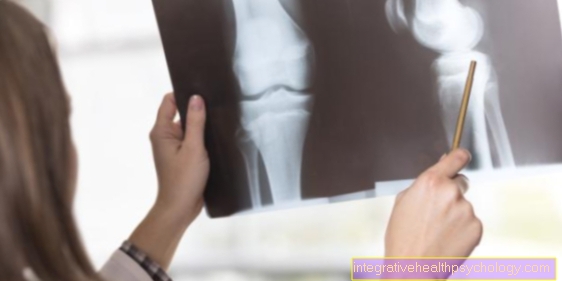
The Diagnosis is primarily done by the doctor by taking the previous history - anamnesis, a functional examination, possibly roentgen or MRI.
You can find medical therapy under the topic Impingement Syndrome. An interdisciplinary collaboration with the physiotherapist and the shared view of the X-rays are desirable and will significantly improve the efficiency of physiotherapy treatment.
Impingement syndrome: prognosis
The earlier the consistent medical and physiotherapeutic treatment of the impingement syndrome is started, the faster the treatment success and the chance of a complete healing of the problem. In the majority of those affected, a significant improvement in terms of symptom relief and functional improvement occurs over the course of several weeks, which can be achieved even with advanced disease. In addition to medication and passive physiotherapeutic techniques, the success of the treatment depends on the intensive active cooperation of those affected.
However, it can happen that an impingement syndrome believed to have been resolved occurs again after a long period of time due to resumed or unknown exposure (sport, gardening, heavy physical work at work) (relapse).
In order to avoid a relapse, it is advisable to continue the exercises for 2-3 months even when the symptoms are free.
If, in the long run, conservative therapy (medical treatment options without surgery) cannot achieve freedom from pain under stress, surgical treatment should be considered, which in most cases has a good chance of success in the long term. However, in the event of a relapse, the previous level of performance cannot always be achieved even with surgery.
Figure impingement syndrome

Impingement syndrome (shoulder)
(Narrowing, crushing of the tendons)
- Collarbone - Clavicle
- Upper bone muscle -
Supraspinatus muscle - Raven beak collarbone ligament -
Coracoclavicular ligament - Shoulder and collarbone
steer -
Articulatio acromioclavicularis - Raven beak shoulder band -
Coracoacromiale ligament - Shoulder corner - Acromion
- Bursa -
Subacromial bursa - Supraspinatus tendon
(Upper bone tendon) - Tendon of the long biceps head
- Upper arm shaft - Corpus humeri
- Two-headed upper arm muscle (biceps),
long head -
Biceps brachii muscle, caput longum - Humerus head -
Caput humeri - Raven beak process -
Coracoid process - Shoulder blade - Scapula
You can find an overview of all Dr-Gumpert images at: medical illustrations


.jpg)