Pulmonary blood flow
Synonyms in the broader sense
Lungs, alveoli, bronchi
Medical: Pulmo
English: breathing organ, lungs
Pulmonary blood flow
With pulmonary blood flow, the lungs are supplied by two functionally different vessels that come from the small and large body circulation.
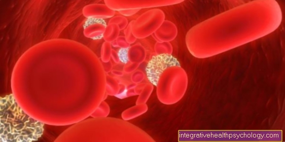
The vessels of the small circulation (pulmonary circulation) transport the entire volume of blood in the body through the lungs in order to absorb new oxygen. They are in the service of the entire body and are also called Vasa publica (public vessels).
The vessels of the great circulation (body circulation) in the pulmonary blood flow are only responsible for the oxygen supply of the lung tissue. Therefore they are also called Vasa privata (own vessels).
All of the following characteristics relate to the flow of blood in the vessels of the small circuit, which are much more important for the functioning of the lungs.
Basically, it must be said that the pulmonary blood flow to the pulmonary vessels is not controlled, as is so often the case, on the basis of the prevailing blood pressure. That makes sense when you consider that the incoming blood should be available again for the large circulation as quickly as possible.
Instead, another mechanism is used for regulation: hypoxic vasoconstriction. This means that the amount of oxygen in the alveoli determines the amount of blood flow.
The more oxygen, the more blood flows through this section; the less oxygen (hypoxia) the less blood (vasoconstriction). This mechanism is mediated by proteins in the cell wall (potassium ion channels) of the alveoli, which change their shape when the oxygen content rises and thus cause a contraction, i.e. contraction. Can introduce constrictions of the vessels.
Assume that an air-conducting section is completely clogged with a foreign object. As a result, fresh air can no longer get into the alveoli. The blood flowing through these alveoli could not take in fresh oxygen. This old blood would still be pumped through the body without transporting oxygen. This scenario is avoided by hypoxic vasoconstriction.
The blood pressure in the pulmonary vessels is low (only ¼ of the pressure in the main artery (aorta)). This prevents the high pressure from forcing liquid from the smallest blood vessels (capillaries) into the alveoli. If this does happen, fluid collects in the lungs (pulmonary edema).
Common causes of pulmonary edema are pumping weakness of the left heart (left heart failure), an increased amount of blood, pneumonia caused by pathogens or an occlusion of a larger vessel in the lungs (pulmonary embolism).
The risk of pulmonary edema is an increase in the distance for the gas to be exchanged from the alveoli during the pulmonary blood flow into the vessels and back. The leading sign of pulmonary edema is shortness of breath (Dyspnea).
Anatomy of air ducts

- Right lung -
Pulmodexter - Left lung -
Pulmo sinister - Nasal cavity - Cavitas nasi
- Oral cavity - Cavitas oris
- Throat - Pharynx
- Larynx - larynx
- Windpipe (approx. 20 cm) - Trachea
- Bifurcation of the windpipe -
Bifurcatio tracheae - Right main bronchus -
Bronchus principalis dexter - Left main bronchus -
Bronchus principalis sinister - Lung tip - Apex pulmonis
- Upper lobe - Superior lobe
- Inclined lung cleft -
Fissura obliqua - Lower lobe -
Inferior lobe - Lower edge of the lung -
Margo inferior - Middle lobe -
Lobe medius
(only on the right lung) - Horizontal cleft lung
(between upper and middle lobes on the right) -
Horizontal fissure
You can find an overview of all Dr-Gumpert images at: medical illustrations

- Bronchiole
(cartilage-free smaller
Bronchus) -
Bronchiolus - Branch of the pulmonary artery -
Pulmonary artery - End bronchiole -
Respiratory bronchiolus - Alveolar duct -
Alveolar duct - Alveolar septum -
Interalveolar septum - Elastic fiber basket
of the alveoli -
Fibrae elasticae - Pulmonary capillary network -
Rete capillare - Branch of a pulmonary vein -
Pulmonary vein
You can find an overview of all Dr-Gumpert images at: medical illustrations


