Mitral valve stenosis
Definition of mitral valve stenosis
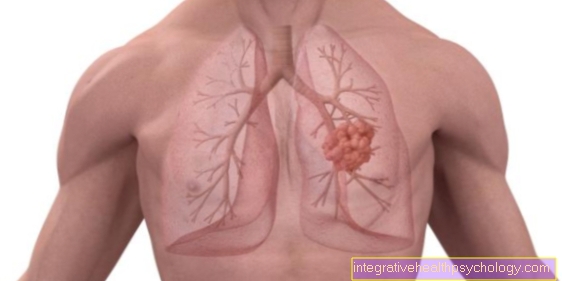
Mitral valve stenosis is a narrowing of the heart valve that separates the left atrium from the left ventricle.
The narrowing of this valve impairs the flow of blood between the left atrium and the left ventricle. The normal opening area of the mitral valve is approx. 4-6 cm2.
Read more about the topic here: Valvular heart disease
If this area is reduced by half or more, it is called mitral valve stenosis. As a result, there is a filling disorder of the left ventricle in the filling phase of the heart's action. Since the left ventricle does not fill up with enough blood, there is a reduced ejection of blood into the body's circulation and there may be an undersupply of vital tissues.
In addition to the decreased ejection into the circulatory system, there is an increase in the pressure difference between the left atrium and the left ventricle. The duck in the left atrium increases and expands it like a water balloon (so-called. Dilation). The expansion / expansion (Dilation) of the left atrium, there may also be a backlog of blood via the pulmonary vessels into the Lungs come (pulmonary hypertension). So the right one must heart work more because it also has to pump against the blood backlog in front of the left atrium.
There is a strain on the right Heart Over a longer period of time, so-called right heart failure can occur. As a result of the expansion of the atrium, some sick sufferers Cardiac arrhythmias, in particular the so-called Atrial fibrillation. The Mitral valve regurgitation is a weakness in the closure of the mitral valve. This means that with every pumping action an additional volume (Regurgitaions volume) is pumped into the atrium, which can also cause the left atrium to overstretch, causing symptoms similar to the Mitral valve stenosis.
Frequency of mitral valve stenosis
The Mitral valve stenosis is one of the most common acquired heart defects and affects women more often than men. The Mitral valve stenosis makes up about 20% of heart valve defects.
Therapy with penicilin decreased the incidence (frequency) of mitral valve stenosis, as with penicillin a successful remedy for grief-positive pathogens such as streptococci had been found.
3-4% of Europeans have heart valve disease. In patients with mitral valve stenosis, the 5-year survival rate is approximately 45-80%. About half of the patients with mitral valve stenosis and about 20-30% with Mitral valve regurgitation develop arrhythmias.
history
The history of mitral valve stenosis is essentially limited to the new surgical intervention methods such as balloon dilatation.
Causes of Mitral Valve Stenosis
The causes of mitral valve stenosis are mostly infectious Origin, but can also be innate.
The rheumatic fever plays a crucial role in acquired heart valve defects. The rheumatic fever is triggered by group A streptococci and often affects the lining of the heart.
This can cause the inner lining of the heart to become inflamed, which can lead to so-called cardiac inflammation (Endocarditis) come. In the course of the infection, this spreads to the mitral valve, as this also consists of the tissue of the inner lining of the heart. The valve defect can occur 20-30 years after the rheumatic fever.
In acute (sudden) rheumatic fever, up to 50% of patients can suffer from a heart valve defect. The consequences of the inflammatory reaction of the streptococci are calcification of the mitral valve and the resulting tightness and restricted mobility.
The Mitral regurgitation is often the result of an inflammatory-degenerative process or a past heart attack. Heart parts can be damaged here (Papillary muscles, papillary tendons), which stabilize the valve apparatus and support it in its opening. If these parts are damaged, the cusps of the mitral valve can protrude into the atrium during the pumping action of the heart (Mitral valve prolapse). The inadequate valve can be imagined as a faulty check valve in which the blood is pumped in the wrong direction during the pumping action. Generally speaking, the causes of mitral regurgitation can be found in organic (primary) and functional (secondary) Classify causes. Organic causes, such as infectious processes, lead almost directly to damage to the mitral valve, while a functional cause is an underlying disease that causes damage to the mitral valve.
Symptoms
The main or guiding symptom of mitral valve stenosis or the Mitral regurgitation is the shortness of breath (medical: Dyspnea). The shortness of breath is caused by the backflow of blood into the lungs.
Through this backlog in the lung, the liquid part of the blood is pressed out into the lung tissue and thus makes the transport of oxygen into the blood more difficult.
The reduced oxygen transport makes breathing difficult or shortness of breath. Most of the time, the shortness of breath only occurs when the patient is exerted, as the heart works more and the congestion in front of the left ventricle is increased. If the constriction is particularly difficult, the shortness of breath can occur even when the patient is resting. Another consequence of backlogging in the lungs can be seizures Coughing up blood (medical: Hemoptysis) be. Here the congestion in the lungs increases and the solid constituents of the blood (red blood cells) also leak into the lungs, causing the sputum to turn red
With long-lasting Mitral stenosis the heart can change as a result of the increase in pressure in the left atrium. The expansion of the left atrium can lead to the so-called Atrial fibrillation to lead.
With atrial fibrillation, the blood flow (hemodynamics) is disturbed and blood clots can form, which can spread into the body and cause further clinical symptoms. The stress on the right heart manifests itself in a backlog of blood in front of the right heart.
This backlog can lead to a Enlargement of the liver and can cause water to build up in the legs (Leg edema). Due to the reduced ejection volume (with reduced filling of the left ventricle), some patients suffer from one peripheral cyanosis (Blue discoloration of the skin). This is caused by the increased exhaustion of oxygen from the blood.
diagnosis

The diagnosis of mitral valve stenosis is made in most cases after an interview with the patient (anamnese), in which the patient presents his symptoms. In the case of mitral valve stenosis, the patient notices, for example, a reduced resilience in everyday situations and / or symptoms from the above paragraph.
If mitral valve stenosis is suspected, the doctor will want to confirm his train of thought with a physical examination. This is usually done first with the stethoscope. When examining the left half of the chest in the fourth intercostal space (medical: 4th Intercostal spacehear abnormal heart murmurs.
Additional diagnostics would be the writing of an EKG in which the doctor can record the electrical activity of the heart. Here the doctor could recognize signs of atrial fibrillation (restless baseline in the ECG) or signs of cardiac stress.
The doctor can also use imaging techniques to support his diagnosis. The echocardiogram enables the doctor to make an ultrasound image of the narrowed mitral valve in order to make a statement about the extent to which the valve is narrowed. Since the echocardiogram can also record the flow of blood over the valve, this examination is considered crucial in the diagnosis of mitral valve stenosis.
Another possibility of examination using ultrasound is the so-called swallow echo. The anatomical proximity of the heart to the esophagus is exploited in that the ultrasound head is swallowed by the patient. In this way, the functioning of the heart valves can be assessed and mitral valve stenosis diagnosed.
Further imaging such as x-rays, computed tomography and MRI can provide information about the load on the heart and changes in the heart structure and valve architecture. However, compared to the echocardiogram, these methods are more expensive or have a high level of radiation exposure.
How is mitral valve stenosis treated?

The therapy of Mitral valve stenosis can be done either conservatively or surgically. The conservative therapy of mitral valve stenosis is mostly a drug therapy of the volume load on the heart caused by the defective mitral valve.
The task of the drugs is to reduce the volume of blood that is in front of the broken one Valve (Heart valve) to reduce congestion in order to relieve the heart.
In general, the Heart work (Heart rate X stroke volume) as an increase in the work of the heart increases the symptoms of mitral valve stenosis / mitral valve regurgitation. The means of choice for a therapy for mitral valve stenosis is here Diuretics (Drainer). Dehydrators reduce the blood volume slightly and thus also reduce the stroke volume. If, in addition to the symptoms, there is pulmonary hypertension, vasodilators can also be used in therapy that Blood pressure reduce. If other severe symptoms of mitral valve stenosis occur, these must also be treated with medication. At Atrial fibrillation need for example blood thinners and Beta blockers be used to risk the embolism and the Heart rate to reduce.
Sometimes with a conservative therapy of mitral valve stenosis insufficient therapeutic result can be achieved. The indication for surgical therapy depends on the patient's symptoms and cardiac function. If the heart function is impaired, for example if the blood expectorates below 60%, surgical treatment of mitral valve stenosis would be possible.
Surgical therapy includes several methods to restore or expand the, for example, narrowed mitral valve. The balloon dilatation (percutaneous balloon mitral valvuloplasty) is a method in which a catheter is used to insert a small balloon over the groin in the area of the mitral valve.
This procedure is particularly gentle on the patient, since the Rib cage does not have to be opened. By inflating the balloon, the constricted Mitral valve expands and thus increases blood flow between the left atrium and ventricle. In addition, a so-called commisurotomy can be carried out, in which the calcified valve tissue is removed and a functional valve can be produced. Valve reconstruction is often performed with the inadequate mitral valve and has a lower post-operative mortality compared to valve replacement. If these surgical procedures are insufficient or cannot be performed, an artificial valve can be used. This can be artificial or can come from a biological preparation (pig, human).
Artificial heart lobes need long-term therapy of blood thinners and Aggregation inhibitors be treated.
rehabilitation
The rehabilitation of the Cardiovascular system is a broad field in itself.Depending on the underlying disease, different methods are of course chosen and different goals are aimed for. The mitral regurgitation or the Mitral valve stenosis is one of the heart valve diseases in the rehabilitation sector.
It is recommended to take part in a regular and controlled training program, in which the program is adjusted to the respective pump function of the heart patient. This allows the heart muscles to strengthen and can therefore endurance win.
With prosthetic mitral valves, prolonged physical stress should be avoided, as the high pulse frequency puts too much strain on the valve. The frequency can also be reduced here with medication (under 80 per minute) for example through beta-blockers.
prophylaxis
The prevention of Mitral valve stenosis largely takes place in avoiding general risk factors for cardiovascular disease.
This mainly means that one should prevent the secondary causes of mitral valve diseases (Heart attack, diabetes...) Here, for example, the focus is on a healthy diet.
Unhealthy nutrition can lead to secondary diseases such as diabetes, which increase the risk of heart disease. Smoke is playing an increasingly important role in the prevention of cardiovascular diseases.
The poisonous substances in cigarettes lead to stiffening and damage to the vessel walls, and thus disrupt the flow of blood. Excessive alcohol consumption also damages the blood vessels. Regular exercise is one of the most effective methods of preventing the secondary causes of mitral valve disease. In order to prevent other diseases such as inflammation of the inner lining of the heart and the formation of blood clots, prophylaxis with medication is also required. These would be for example Antibiotics and blood thinners. Of the Blood pressure In the broader sense, it must also be kept stable with medication, as excessively high blood pressure could damage the mitral valve further and thus worsen the clinical picture. Lowering the blood pressure also ensures that the heart is relieved sufficiently.
forecast
An untreated mitral regurgitation or a Mitral valve stenosis would certainly lead to premature death.
This is of course different for each patient, especially because the mitral valve diseases usually progress slowly until they become clinically noticeable. The heart changes functionally and anatomically to adapt to the diseased valve. As I said, this works differently well or badly for each person with the disease.
In patients requiring surgery, the 8-year survival rate is 89%. The prognosis essentially depends on the pumping capacity of the heart. In patients who had normal pumping function, the 10-year survival rate is approximately 72%, and in patients who have impaired pumping function, the 10-year survival rate is 32%. Sudden deaths are relatively rare, with an incidence of around 0.8%.
Summary
Mitral valve diseases (mitral regurgitation and mitral valve stenosis) are among the slowly progressive diseases.
They often take years to manifest themselves clinically and are often associated with bacterial infections and degenerative processes. In the long term, the mitral valve diseases lead to a reduced pumping capacity of the heart, which often manifests itself in the clinical appearance of shortness of breath and reduced resilience.
Mitral valve disease can often be medicated with Antihypertensive, Frequency sink and Drainage Preparations are treated. However, if an operative procedure is necessary, it aims to repair the damaged valve or valve apparatus, or even to replace it completely. Which procedure would be most suitable is usually decided after consultation with the respective cardiologist or heart surgeon.
The 10 year survival rate is very good with today's surgical procedures, and enables the patient to participate in their everyday life again without hindrance. In very few cases, sudden death from mitral valve disease can occur.