Middle ear
Synonyms
Latin: auris media
English: middle ear
anatomical: Tympanic cavity (Cavitas tympani)
introduction
The middle ear is a space filled with air that is lined with mucous membrane and is located in the petrous bone of the skull. The ossicles are located in it, through which the sound or the vibration energy of the sound is transmitted from the external auditory canal via the eardrum and finally to the inner ear.
anatomy

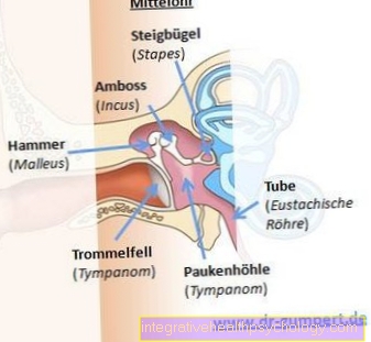
Anatomically, the ossicular chain, consisting of, becomes the middle ear hammer (lat. Malleus), anvil (lat. Incus) and stirrup (lat. Stapes), counted. They are articulated together. The hammer is adjacent to the eardrum (Membrana tympani), which represents the boundary between the outer ear and the middle ear. The anvil is attached to the hammer and is in turn connected to the stapes in the middle ear. The latter ends with its stirrup footplate at the oval window (Fenestra vestibuli).
The ossicles in the middle ear are the smallest boils in the human body and, in addition to transmitting sound, also perform the function of amplifying sound by a factor of 1.3. This is achieved through the leverage of the ossicles. Overall, the movements of the ankle chain are pendulum movements and their mobility is influenced by two muscles: Tensor tympani muscle („Eardrum tensioner") and Stapedius muscle (starts at the stirrup). Both muscles reflexively reduce the transmission of sound in the case of loud sound stimuli and thus fulfill a certain protective function. When the M. tensor tympani the eardrum tightens in the middle ear; with contraction of the M. stapedius the sound conduction chain is stiffened and the sound transmission to the inner ear is reduced. This filter function should be particularly important for high notes ("High pass filter“).
Figure ear

A - outer ear - Auris externa
B - middle ear - Auris media
C - inner ear - Auris interna
- Ear strip - Helix
- Counter bar - Antihelix
- Auricle - Auricula
- Ear corner - Tragus
- Earlobe -
Lobulus auriculae - External ear canal -
Meatus acousticus externus - Temporal bone - Temporal bone
- Eardrum -
Tympanic membrane - Stirrups - Stapes
- Eustachian tube (tube) -
Tuba auditiva - Slug - Cochlea
- Auditory nerve - Cochlear nerve
- Equilibrium nerve -
Vestibular nerve - Inner ear canal -
Meatus acousticus internus - Enlargement (ampoule)
of the posterior semicircular canal -
Ampulla membranacea posterior - Archway -
Semicircural duct - Anvil - Incus
- Hammer - Malleus
- Tympanic cavity -
Cavitas tympani
You can find an overview of all Dr-Gumpert images at: medical illustrations
The tympanic cavity in the middle ear is bounded by several walls. The side wall (Paries membranaceus) represents the limit to outer ear It is mainly formed by the eardrum. The inner wall (Paries labyrinthicus) is the border to the inner ear. A protrusion is particularly noticeable here; the so-called Promontory. It is the basal spiral of the Inner ear. The lower wall (Paries jugularis) forms the floor of the tympanic cavity. Via the back wall in the middle ear (Paries mastoideus) one reaches further air-filled cells via a corridor (Cellulae mastoideae) of the petrous bone. A Otitis media spread because there is a direct connection. The roof of the tympanic cavity limits the upper wall (Paries tegmentalis). Another important opening or connection of the middle ear includes the anterior wall (Paries caroticus) - the ear trumpet opening. The ear trumpet (Tuba auditiva) in the middle ear provides an open connection between the middle ear and throat here. It consists of one third of bony and two thirds of cartilaginous material. The corporeal part follows the bony part located in the petrous bone and widens towards the throat like a trumpet like a funnel. The tube guarantees constant ventilation of the middle ear and opens with every swallowing act. This results in a pressure equalization between the air pressure in the middle ear and the environment. That is why it is often advisable to suck candy or swallow frequently during flights in order to avoid “pressure on the ears”. The eustachian tube has a special surface with cilia as an additional protective measure, which is supposed to keep germs away from the middle ear by beating towards the throat. If this system fails, it can lead to ascending otitis media caused by bacteria.
The neighborly relationships are of clinical importance, especially in diseases of the middle ear, since from here a severe purulent inflammation can spread to adjacent spaces. Can follow a Meningitis, Brain abscessesInflammation of the mastoid process of the temporal bone (Mastoiditis), Visual disturbances and paralysis of the Facial muscles.
Another anatomically important structure runs directly through the middle ear, protected only by a fold of the mucous membrane. It is a small nerve (Chorda tympani), which is responsible for the sensation of taste. At a Otitis media this nerve can be affected. Affected people report taste disturbances and reduced salivation.
Function of the middle ear

In addition to "simple" sound transmission, the most important task of the middle ear is the so-called sound wave resistance adjustment (Impedance) represent.
The incoming sound reaches the eardrum via the external auditory canal. If the inner ear filled with fluid were to connect directly, approx. 99% of the sound waves would be reflected because the sound wave resistance between air and inner ear fluid is too great. This problem is circumvented with the help of the middle ear. The sound energy is effectively brought to the oval window via a hammer, anvil and stirrup. Two mechanisms for impedance matching are important here. First, as already mentioned, the ossicles cause an increase in pressure on the oval window through different lever arms. However, the second effect takes over the significantly larger part of the adjustment processes. The principle here is the surface effect between the eardrum and the oval window. Since the eardrum is approx. 17 times larger than the oval window, an equal force must be distributed over a smaller area. This results in an enormous increase in sound pressure by a factor of 30.
Overall, the middle ear and its impedance adjustment reduce sound reflection to 35%, which increases hearing by 10-20 decibels (dB) depending on the frequency.
Summary
The middle ear represents an indispensable part of hearing. In diseases such as otitis media, this is sometimes severely restricted. Complications complicate the clinical picture.


