MRI of the lumbar spine
introduction
Magnetic resonance imaging, also known as MRI for short, is an imaging procedure in medicine. The process works without harmful ionizing radiation. In the clinic it is used to make sectional images of the body. During the examination, the magnetic resonance tomograph generates a magnetic field and alternating magnetic fields, whereby atomic nuclei, in particular hydrogen nuclei (Protons), are set in motion in the cells of our body.

Creation of the sectional images
Electrical signals are generated that can be registered by means of a receiver circuit.
The formation of contrasts between different structures and tissues is based on the relaxation time and the proportion of water content of the different cell types. Ultimately, we measure the proportion of hydrogen atoms in the tissue structures. The types of tissue differ from one another due to the different proportions of hydrogen atoms.
Due to the good contrast, organs, different tissue and soft parts can be displayed very well and offer better imaging than conventional X-rays. The spinal cord and nerves, nerve water (Liquor), Intervertebral discs (including herniated disc), facet joint and ligaments of the spine.
Read more on the subject at: MRI of a herniated disc
Procedure of an MRI
preparation
Patients are informed of the exact procedure before the examination and must also sign a written information and consent form. If you have any further questions, please contact the trained staff or the treating doctor.
For the examination itself, no further preparatory measures, such as laxative measures (this is not necessary for an MRI of the lumbar spine, but it is necessary for an MRI examination of the small intestine) the day before.
Patients are asked to remove their clothing for the examination. In particular, metallic objects such as jewelry, watches, piercings, hairbands and underwear with metal trimmings should be removed because they are attracted by the magnetic field and therefore pose a risk of injury.
You may also be interested in this topic: Clothing in the MRI - what should I wear?
Procedure of the investigation
Lying on the table, the patient is finally covered with a blanket and driven into the MRT examination device. Patients are informed in advance that the magnetic resonance tomograph is very narrow and very loud. In consultation with the attending physician, agitated and anxious patients can be given a sedative in advance to reduce anxiety. Patients with claustrophobia should express this beforehand and, if necessary, find out about the alternatives.
In order to produce very good pictures, interference caused by movement should also be avoided at all costs. To protect against the volume and the various knocking noises, the patient is given headphones. The patient should lie relaxed and comfortable during the examination.
Read more on this topic at: The procedure of an MRI and MRI for claustrophobia
Duration of the investigation
The duration of the examination is around 15-25 minutes.
In addition come possible preparations such as undressing the patient, positioning them on the examination table and then evaluating the images taken.
Some findings require some time to be evaluated and to make a diagnosis with certainty. You can also longer waiting times arise because the MRI examination is also often used in emergencies.
Is a contrast agent necessary?
Contrast media are not used in medicine for therapy, but are used to advance and simplify the detection of diseases. They are used in imaging procedures, including MRI.
While iodine-containing control agents are used in conventional X-ray diagnostics, a distinction is made between two different substances in the MRI of the lumbar spine.
The extracellular contrast agents are Gadolinium-containing compounds that are easily excreted by the kidneys. The gadolinium it contains causes faster magnetization of water in its immediate vicinity. For example, specific vessels can be displayed very brightly. Intracellular contrast media, on the other hand, accumulate in certain tissues. For example, they are used to represent the liver and pancreas (pancreas) used.
Contrast media are used when they are supposed to help visualize soft tissue better. The images are richer in contrast and any abnormalities can be better recognized. The MRT contrast media have the advantage that they are very well tolerated. They can also be used if the patient already has a known iodine allergy or hypersensitivity reactions to medical devices occur frequently.
Nevertheless, complications can also arise with the MRI contrast media used. In very rare cases it can also develop into a nephrogenic systemic fibrosis to lead. It is a disease of the connective tissue and the skin. It can lead to a decrease in movement as the joint stiffens. In the further course, damage to the internal organs can ultimately also occur. Patients with severe kidney disease have an increased risk of developing it. If one of the following symptoms should occur several hours or even days after the MRI of the lumbar spine, a doctor should be consulted immediately to clarify a late complication.
- itching
- Rash
- Pain
- nausea
Read more on the subject at: Contrast media or MRI with contrast agent
Indications
There can be a number of reasons why you need to have magnetic resonance imaging (MRI) of the lumbar spine (lumbar spine). Often the MRI examination is not the first choice because it takes much longer than computed tomography (CT) and is associated with considerably higher energy and cost. The advantages of an MRI, however, are the better representation of the soft tissues and vessels. In the area of the lumbar spine, this means that the intervertebral discs and the spinal cord as well as the blood vessels are shown very well and in detail on the MRI images, so that a good diagnosis is possible.
In addition, an MRI scan is preferable in young patients and especially in pregnant women, as there is no radiation exposure there like with X-rays or CT.
Since cross-sectional images are made of the examined area during an MRI examination, it is possible for the radiologist to assess all areas and levels of the lumbar spine.
An MRI is indicated for diagnosing congenital malformations in the lumbar spine and if bony changes are suspected due to an accident. This allows the extent of the injury or deformity to be estimated and possible effects on the spinal cord to be identified. Often, however, the MRI is a follow-up examination that follows the X-ray or CT.
An MRI is indicated for chronic back pain in the lumbar spine. The intervertebral discs are well represented and a herniated disc or a protruding disc can be seen there.
In contrast to CT, MRI has the advantage that the affected spinal cord segments can often be better recognized. MRI is also suitable for detecting or excluding tumors or inflammation in the spinal column area. It is often necessary to carry out the examination with a contrast medium. A lumbar spine MRI is also used to assess the success of the operation, as well as to monitor the progress of multiple sclerosis (MS). If there is a fracture in the vertebra, an MRI is often done to determine whether the fracture is affecting the spinal cord or blood vessels, or whether there is bleeding in addition to the fracture.
In the case of a navel piercing, performing an MRI of the lumbar spine is limited.
You can find out more about the topic here: MRI and piercings, MRI for a herniated disc such as MRI in multiple sclerosis
Contraindications
An MRI scan is contraindicated in a patient with a pacemaker due to the magnetic fields. The magnetic field would disrupt the function of the pacemaker and put the patient at considerable risk. Furthermore, the examination cannot be carried out on patients who have metallic foreign bodies such as prostheses in their bodies. In such a case, the examination cannot be carried out.
Cost of an MRI of the lumbar spine
The cost of an MRI scan of the lumbar spine can vary widely. They are directed e.g. according to insurance (GKV or private health insurance), number of shifts or administration of contrast media. If there is a medical indication for an MRI examination, such as a referral from a doctor, both statutory and private health insurances usually cover the costs of the examination. However, if the examination is only carried out at the request of the patient, the costs must be borne by yourself. The costs for an MRI of the lumbar spine are between € 400 and € 800.
Read more on this topic: Cost of an MRI examination
MRI for a herniated disc
In the case of a herniated disc (in medical terminology as Disc prolapse or Nucleus pulposus prolapse The inner core of the intervertebral disc falls out, causing painful or neurological symptoms. Various imaging techniques can be used to confirm the diagnosis.
In addition to computed tomography, the MRI of the spine is also used here, as this examination can show the structures of the tissue and nerves particularly well.
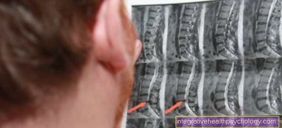
Many images of the entire spinal column are taken so that a herniated disc in the lumbar spine can be easily recognized. In the MRI, the various structures of the lumbar spine, such as the vertebral body, spinal cord, and nerve water, appear differently depending on the weighting (T1, T2, PT). Therefore, knowledge of the physical parameters is always necessary for diagnosis. It is particularly important to show the intervertebral discs with the gelatinous nucleus in the center of the intervertebral disc, which is demarcated in the MRI. That gelatinous core, too Nucleus pulposus called, is surrounded by a fiber ring. If the fibrous ring tears when it is overloaded, the gelatinous nucleus can advance backwards into the spinal cord. The spinal cord lies behind the vertebral bodies and can be recognized on an MRI by its light, fibrous color. If there is such a herniated disc, the advancement of intervertebral disc tissue (Jelly) in the spinal canal. The spinal cord is also significantly narrowed at this point. In addition, you can see a smaller distance between the vertebral bodies that are adjacent to the affected intervertebral disc.
The diagnosis is made by a radiologist and passed on to the treating doctor
Read more on the topic: Herniated disc of the lumbar spine
MRI in tumor diagnosis of the lumbar spine
MRI is a very important and frequently used imaging method in the diagnosis of lumbar spine tumors. Since the MRT can very well show different soft tissue qualities of different types of tissue, it is used to exclude tumors or to assess existing tumors with regard to their location and size. Especially before surgical interventions, knowledge of the location and relationship to other surrounding structures is extremely important for the surgeon and for the choice of subsequent treatment. Here, too, the advantage of MRI is the lack of radiation exposure compared to conventional CT and X-rays, and tumors can also be performed very well with the aid of MRI without the administration of contrast media.
The MRI images taken can be used to make an initial assessment of the tumor with regard to its degeneration, i.e. whether it is a benign (benign) or malicious (malignant) Tumor.
Malignant tumors are noticeable on MRI because of their invasive and aggressive growth. They often grow quickly, displacing the surrounding tissue. Another easily observable indication of a malignant tumor can be the presence of newly formed vessels near the tumor, since malignant tumors are able to produce substances that stimulate the surrounding vessels to grow.
Benign tumors, on the other hand, are characterized by a clear demarcation from the neighboring tissue, and their growth is significantly less aggressive and also slower.
Nevertheless, the findings should always be confirmed histologically, for example by an intraoperative biopsy. Primary malignant new growths are rare in the lumbar spine area. Rather, one finds metastases (daughter tumors) in the vertebral bodies in the MRI of the lumbar spine. Metastases from breast cancer, prostate cancer or a lung tumor are particularly common (Bronchial carcinoma).
Sacroiliac joint (SIJ) MRI
The sacroiliac joint (ISG) can be affected by misalignments and blockages due to its high mechanical stress. These can lead to ISG syndrome. It is characterized by a mixed form of lumbar and sacral pain.
Often the patients also report diffuse pain in the area of the hip and the deep lumbar spine (lumbar spine). The pain gets worse with exercise and when sitting, but can also occur spontaneously and just as spontaneously go away again. In addition to the clinical examination, which includes the functional tests of the hip, an MRI of the lumbar spine can also be used to diagnose an ISG syndrome. This is particularly useful if the presence of a rheumatic disease is to be excluded. In particular, the inflammation of the sacroiliac joint, which is medically referred to as sacroiliitis, can be safely depicted in an MRI. The articulated structure of the sacroiliac joint can be assessed very precisely in the MRI. Adhesions or acute inflammatory processes can also be diagnosed by the examination.
Also read more on the topic: ISG blockage
MRI for cysts
A cyst is a fluid-filled cavity that can be found in different tissues. There are often cysts in the breast, in the ovaries (see ovarian cyst), in the head or in the kidneys. The fluid can be blood, pus, sebum, or tissue fluid that is then enclosed in a thin or tough capsule. In most cases, cysts are benign changes, but degeneration can also occur here. They also come in different sizes and can appear at any age. Most of the time, the cysts cause no symptoms and are discovered by chance during a routine examination. Symptoms of cysts on the lumbar spine can occur when the size of the cyst impairs the function of important structures such as nerves or vessels.
In an MRI of the lumbar spine, facet joint cysts are particularly common, which are usually signs of wear on the facet joint (Facet joint arthrosis) represents. More rarely, arachnoid cysts emanating from the meninges are found in an MRI of the lumbar spine. Therapy, such as removing or puncturing the cyst, is often only performed when such symptoms appear, or at the patient's request. Using the MRI of the lumbar spine, it can be ensured that it is only a benign mass, for example. In addition, the size and the positional relationships can be precisely determined. This makes it easier to choose the right therapy in the following.

