MRI of the brain
introduction
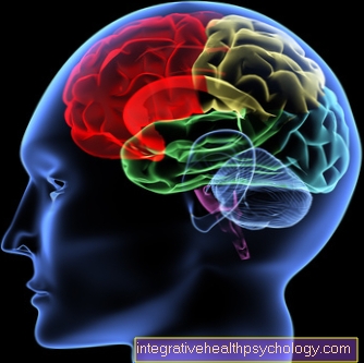
MRI imaging of the brain is used for many different questions and, in addition to CT imaging, it is another way of getting a detailed representation of the skull and brain tissue.
The MRT is particularly well suited for the display of soft tissue, while with a CT the bone display is better.

The indications for an MRI scan of the brain include the diagnosis of a stroke or the precursors of a stroke, masses such as benign or malignant brain tumors, water retention, etc., so-called demarking brain diseases such as multiple sclerosis, so-called degenerative brain diseases, such as the various forms of dementia or Parkinson's, severe headaches (e.g. migraines), epilepsy or birth defects.
The MRI can be used both for the initial diagnosis, as well as for monitoring the progress, for therapy planning or for therapy control.
Read more on the subject at: MRI of the head
Do you need contrast media?
Whether a contrast agent is required or used in the context of an MRT examination depends on the question - i.e. on the structures that are to be examined with particular attention. Since MRI images are displayed in black and white and the range of gray levels is limited, it may be difficult to distinguish between different structures or tissues.
If a contrast agent is given - usually via the arm vein - it can be easier to differentiate between specific tissues and their surroundings. The reason for this is that the contrast agent used in MRI is distributed particularly in the blood vessel system and floods in tissues such as tumors or metastases as well as in tissues that are inflammatory. Thus, e.g. Brain aneurysms, bleeding, foci of inflammation or brain tumors / metastases can be better represented and highlighted by the administration of contrast medium.
The examining radiologist decides whether a contrast agent is used before or during the examination.
Read more on this topic at: MRI with contrast agent.
MRI of the brain in MS
Magnetic resonance tomography (MRI) is used in the context of multiple sclerosis (MS for short) on the one hand to establish a diagnosis in the event of suspicion and, on the other hand, to monitor the progression of an already established diagnosis.
What the MRI image of the brain can show in relation to an MS disease are, in particular, the foci of inflammation that are characteristic of this neurological disease of the central nervous system. The foci of inflammation arise when the body's own immune system falsely recognizes and fights certain structures of the nerves or nerve cells as foreign (so-called autoimmune reaction), so that an inflammatory reaction occurs (also known as "foci of demyelinating").
These foci of inflammation are mainly found in the lateral brain water chambers (periventricular) and in the so-called "bar", a part of the brain that connects the two halves of the brain.In the MRI they usually appear lighter than the surrounding tissue, especially when contrast media is given as part of the MRI diagnosis.
Read more on this topic at: MRI in multiple sclerosis.
MRI of the brain during a stroke
Depending on the cause of the stroke that has occurred, different features can be seen in the MRI.
The MRI is considered to be more accurate and reliable than the CT, as it can also detect particularly small stroke foci. The only disadvantages are the significantly higher cost factor and the longer time it takes to take the image, which is out of the question in an acute emergency.
A distinction can be made between a "hemorrhagic" stroke, i.e. a loss of brain tissue due to bleeding from a ruptured brain vessel, and between an "ischemic" stroke, in which the brain tissue has died due to a reduced blood flow caused by a clogged, closed brain vessel (blocked by E.g. a blood clot = thrombus, embolus).
"Bloody" areas of the brain appear lighter in a contrast agent-assisted MRI image than the rest of the healthy area. On the other hand, areas of the brain that have disappeared due to a vascular occlusion appear darker. In addition, a special representation of the brain vessels (magnetic resonance angiography, MRA) can be carried out as part of the MRT examination, so that blocked or ruptured vessels can be mapped and localized.
Read more about the MRI for a stroke.
MRI of the brain in dementia
The MRI is used in the diagnosis of dementia to distinguish whether it is a primary or a secondary dementia.
Primary dementia is an independent type of dementia, such as Alzheimer's dementia. So-called brain tissue atrophies, i.e. a loss of brain substance or a decrease in brain volume, are characteristic of these primary dementias. This can be recognized in the MRI by a cerebral cortex that is too thin, flattened cerebral convolutions, widened and deepened cerebral furrows and cerebral water chambers that appear enlarged.
The MRI is used to differentiate between the primary forms of dementia, which is important for the subsequent treatment. On the other hand, the MRI can also exclude secondary dementias, i.e. dementias that develop as a result of other diseases, such as Tumors, abscesses, water retention, or brain infarctions.
Read more on this topic at: Dementia.
This is how you can recognize pressure marks on the brain
One speaks of increased intracranial pressure when it rises above 15 mmHg. The increased intracranial pressure is caused by the increase in volume within the bony skull.
A CT or MRI is usually done to detect signs of increased intracranial pressure.
A possible sign of intracranial pressure is the expansion of the CSF spaces, for example if there is a CSF outflow disorder. An asymmetry of the liquor spaces can also be an indication of excessive intracranial pressure. The space between the brain stem and the skull should also be considered. A reduction in this space also speaks for an increased intracranial pressure. A final sign of intracranial pressure can be elapsed convolutions on the image. These indicate a swelling of the brain (Brain edema).
Furthermore, a cause for the increased intracranial pressure, such as a tumor or bleeding, can possibly be found in the MRI of the brain.
Read more on this topic at: Increased intracranial pressure
costs
The costs for an MRI examination of the brain are always covered by the health insurance companies if there is an indication for this, i.e. if the examination is medically justified. If this is not the case and the patient would like to have an MRT examination on their own initiative, without there being a medical reason for this, they must pay for the examination themselves.
For patients with statutory health insurance, the costs for a brain MRI are calculated according to the uniform assessment standard (EBM), for private patients, however, according to the fee schedule for doctors (GÖA).
For patients with statutory health insurance, the costs for pure MRI imaging of the brain skull - and thus also of the brain tissue - are € 126.59 (the costs for displaying the facial skull or the skull base for certain questions are both the same amount).
In the case of private patients, a minimum of € 256.46 to a maximum of € 461.64 can be charged for the MRI examination of the skull - depending on the question and the examination effort. In addition to the costs for imaging only, there are usually additional costs for any contrast agent used, advice or certain storage.
Read more on this topic at: Cost of an MRI examination.
Duration
The length of time it takes to perform an MRI scan of the brain depends on how much time the actual image acquisition process takes.
The pure imaging of the brain usually takes 15-20 minutes, although deviations can also occur here. The duration also depends on whether there is still a contrast agent administration via the arm vein or whether there are additional or special images in certain cutting planes if certain questions are to be investigated.
In addition to the total duration of the MR examination of the brain, there is the waiting time and the preparation time before the start of the examination (taking off clothes, positioning the patient, etc.) as well as the final discussion about the results after the examination.
All in all, the actual image acquisition time usually only takes up a small proportion of the duration of an MRI examination of the brain, but the examination procedure can total 1-1.5 hours - however, an exact time can by no means be given reliably.
Read more on the subject at: Duration of various MRI examinations.