MRI - examination of the abdominal organs
introduction
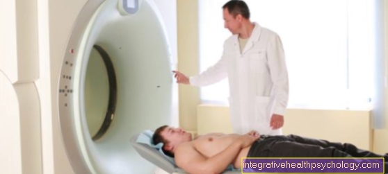
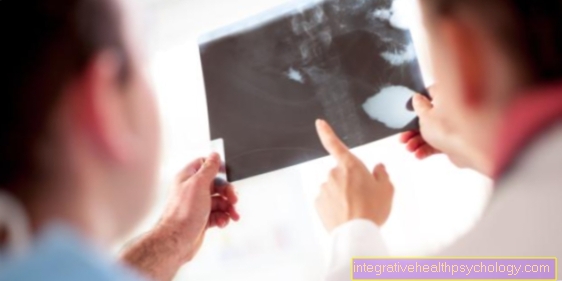
A magnetic resonance tomography is a safe method to get a good insight into the abdominal cavity without having to perform surgical interventions.
An MRI scan of the abdomen (also called an MRT abdomen) is always carried out if other imaging tests have not provided any decisive information about the cause of the discomfort. As a rule, imaging of the abdomen is always necessary when the patient reports symptoms such as pain or chronic diarrhea or when a structure was seen in a previous imaging that could not be assigned and assessed.
Read more on the topic: Procedure of an MRI

Duration of an MRI scan
In addition to the high costs of an MRI examination, a major disadvantage is the length of the treatment. While an X-ray or CT examination only takes a few minutes, an MRI examination can take many times the time. Here too it is crucial which body region is examined.
Investigating a shoulder by an MRI approx. 15 to 25 minutes take the investigation of a Spine however can between 30 and 40 minutes last. One issue that makes many examinations difficult is when patients are under a Claustrophobia Suffer. Due to the tightness of the device, it can sometimes be necessary for a short-acting Sedatives given to the patient until the MRI scan is done. A MRI for claustrophobia is still possible.
Do you have to be sober?
Not for everyone MRI examination you have to be sober. It depends above all on which part of the abdomen is examined with an MRI. If Stomach or intestines must be examined, the patient sober in order to avoid that you can see residues of the food in the MRT, which can lead to overlapping in the final image. Furthermore, an intestine after previous food intake is e.g. always overlaid with air, which can also lead to glare effects in the picture.
When investigating the liver, of the bladder or the Kidneys must be the patient not necessarily sober to be. So it is also possible with normal eating habits to have a MRI of the kidney perform.
If the patient is advised to be sober, at least it will do nothing to eat or drink four hours beforehand. After the MRI examination, the person examined can eat again immediately.
MRI examination with or without contrast agent?
In general, one cannot say that an MRI scan is with or without Contrast media must be carried out. On the one hand, it is crucial what part of the abdomen is examined and, secondly, what exactly the question is. Often a native MRI image is taken first, i.e. the MRI examination without contrast agent is carried out first. If certain structures that are seen cannot be assigned, it may be necessary to MRI with contrast agent to drive.
The contrast agent stains some structures in the body, but leaves others unstained. In case of doubt, the examiner can then identify different tissues in the body.
On the one hand, there is a contrast agent expensive than the native recording, on the other hand too not without risks. It can happen from time to time that the person being examined points to the contrast medium allergic responds. It is therefore necessary to ask the patient beforehand whether an appropriate Contrast agent allergy is known. Another important point is the use of iod containing Contrast media. If the patient is under a Thyroid disease has to switch to another, non-iodine-containing drug.
If there are no objections and the patient has signed the information sheet with the possible risks and side effects, he will receive a venous access placed either in a vein in the back of the hand or in a vein in the crook of the elbow. The patient is then moved into the MRT machine on a couch. If it is decided that the patient must receive a contrast agent in order to improve the representation of the abdominal cavity in the MRI, the contrast agent is applied from the outside via the existing access. The contrast medium floods into the body within a few seconds. The contrast medium is also quickly drained off again, which then has the consequence that the corresponding image of the abdominal cavity becomes indistinct again. So hurry is required. The recordings must then be carried out. You can also repeat the administration of contrast medium, but should apply as little contrast medium as necessary in order to minimize the corresponding risks and dangers of the allergic reaction.
In the following MRI images, contrast agent applications are particularly common. The representation of the Biliary systembecause there are many structures next to each other that have to be differentiated. Investigations of the Intestinesbecause the exact boundary between the intestinal wall and intestinal contents must be shown here (the contrast agent is often drunk beforehand and waited until it has accumulated and distributed in the intestinal tract). The MRI images are then carried out. Also at swallowed contrast media risks and dangers must be observed
Cost of an MRI examination
The MRI provides one of the most expensive diagnostic measures in medicine. The reasons are, on the one hand, the high development costs of the device, the high purchase prices and the high maintenance and servicing costs that are regularly incurred. Furthermore, due to the length of an MRT examination, not as many patients can be examined per day and for this reason the device can generate less profit.
The cost of an MRI scan vary greatly depending on the body region examined. Investigating the shoulder and des Neck area cause the highest cost. These amount to about 600 EUR and can increase with the use of contrast media.
When examining the liver or the Kidneys fall between 350 and 450 EUR on. Here, too, the additional administration of contrast agent can cause costs to skyrocket. When examining the entire colon or small intestine beat costs of approx. 500-700 EUR depending on the length and section of the examination.
The costs for an MRT examination are usually covered by the statutory health insurance. There are some borderline decisions that require an application to be reimbursed first. Of course, the indication given by a doctor is also important. At times, the use of a more or less full-body MRI as a preventive examination has also been discussed. Because of the massive costs, this idea was quickly rejected.
indication
As a rule, after the physical exam still a Ultrasound examination of the abdomen before the indication for an MRI is made. Was e.g. in the ultrasound Gallstone seen and it should be clarified whether there are other stones in the further course of the biliary system, an MRI examination of the abdomen is also recommended.
Even if abnormal structures on the liver were found that could not be correctly assigned, an MRI is indicated. There is an additional option here MRI of the liver to drive.
For some years now there has also been an option instead of one Colonoscopy (Colonoscopy) perform a virtual colonoscopy using an MRI scan. The images of the intestine are recorded in such a way that one can see the intestinal wall and the structures as well as possible Polyps or changes, as seen in a colonoscopy. The colonoscopy is still a lot more precise and is the standard for intestinal examinations.

