annoy
synonym
Nerve cells, neurons, lat .: Nervus, -i
definition
Neurons are nerve cells and therefore part of the nervous system. They serve the
- Admission,
- Processing and
- Forwarding of information.
construction

A nerve cell consists of a cell body (Perikaryon or Soma) and appendages.
There are two types of processes:
- Dendrites and
- Axons.
Read more about the topic here dendrit
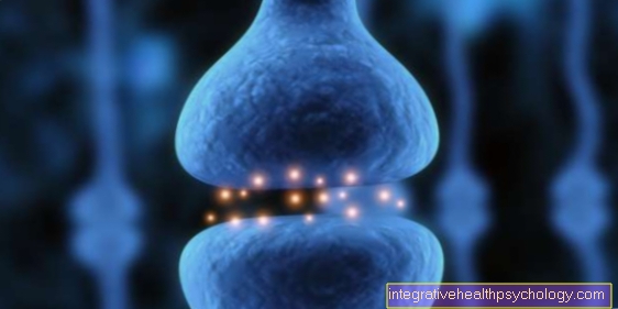
As a rule, a nerve cell has several dendrites. They branch off from the cell body like branches of a tree and this the reception of stimuli. Axons, on the other hand, are used to transmit information over sometimes very long distances of over one meter. Usually a neuron has only one axon. At the axon endings there are numerous synapses that serve to transmit signals from one nerve cell to the next or from the nerve cell to the recipient organ.
One differentiates neurons according to their number of processes:
- pseudounipolar,
- bipolar and
- multipolar.
Multipolar neurons have many dendrites and an axon, while bipolar neurons have a dendrite and an axon. Pseudounipolar axons seem to have only one process, which, however, has dendritic and axonal parts.
Furthermore, one roughly distinguishes:
- sensory of
- motor neurons.
Sensory neurons conduct afferent information.
Afferent means that they receive information in the body's periphery and direct it towards the central nervous system.
For example sensations like:
- Touch or
- Pain.
Efferent neurons, on the other hand - like the motor neurons or motor neurons - carry information that was generated centrally to the periphery and thus trigger a muscle contraction, for example.
One differentiates:
- myelinated (medullary) from
- non-myelinated (marrowless) Neurons.
Myelin is used to isolate a nerve cell and enables a much faster conduction of excitation. For example, myelinated neurons conduct at a speed of around 100 m / s while non-myelinated neurons only conduct at around 1 m / s.
Illustration of a nerve cell

Nerve cell -
Neuron
- Dendrites
- Synapse
(axodendritic) - Cell nucleus -
Nucleolus - Cell bodies -
Nucleus - Axon mounds
- Myelin sheath
- Ranvier lace-up
- Swan cells
- Axon terminals
- Synapse
(axoaxonal)
A - multipolar neuron
B - pseudounipolar neuron
C - bipolar neuron
a - Soma
b - axon
c - synapses
You can find an overview of all Dr-Gumpert images at: medical illustrations
physiology
Information is in the nerves in the form of
- more chemical and
- electrical Activity encoded.
Information is passed on via Action potentials. The basis for this are ion currents.
At the Nerve cell are - in a simplified scheme - the most important ions:
- potassium and
- sodium.
The potassium concentration is in the cell (intracellular) high and outside the cell (extracellular) low, but the sodium concentration is intracellular low and extracellular high.
This ion concentration is primarily controlled by an ion pump, which Sodium-Potassium ATPase reaches the potassium ions in the cell and sodium ions out transported out of the cell.
If the cell membrane were now permeable to sodium and potassium, the ions would flow from the place of high to place of low concentration. Potassium would flow extracellularly, while sodium would flow intracellularly. However, the membrane is not easily permeable to ions, but the permeability is specific channels regulated.
There are channels for Potassium ions and channels for Sodium ions.
The ion current therefore depends on which channels are open and which are closed. In the nerve cells there is a calm - when they are not excited - a Resting membrane potential with clearly negative values:
- about -70 mV.
This resting potential is primarily generated by the constant outflow of potassium ions from the inside of the cell to the outside. This outflow is possible because certain potassium channels are open at rest. If the nerve cell is stimulated, sodium channels in particular open. This causes an influx of positively charged sodium ions, which makes the membrane potential more positive.
If a certain threshold is reached, a Action potential at the top of which the membrane potential assumes positive values:
- about +30 mV.
This is achieved by closing the sodium channels again and reopening the potassium channels, through which potassium ions in turn escape from the cell Membrane potential after this Action potential his again quickly negative resting value.
Excitation conduction
So that the information along the Nerve cell can spread and be transmitted over long distances, must always Action potentials generated along the nerve.
A distinction is made between two types of excitation conduction:
- saltatory and
- continuous Excitation conduction.
In the saltatory conduction, parts of the nerve are so well insulated in regular sections that the excitation here "jump over“Can move from one non-isolated area to the next. These fully isolated areas are called Internodes designated. The short non-isolated area in between will be Ranvier laces called and contain a high number of Ion channelsso that a new action potential is generated here, which can then jump to the next lace ring.
So need a lot less Action potentials than with the continuous Excitation conduction, in which potentials have to be triggered again and again along the entire nerve in closely adjacent sections.
That's why the saltatory Excitation conduction with about 100 m / s much faster than that continuous with about 1 m / s. It only takes place on isolated neurons; the isolation is ensured by myelin, which surrounds the Nerve cell wraps. A pathological demyelination such as that in the Multiple sclerosis (MS) takes place, leads to a significant slowdown in nerve conduction with partial failure of nerve functions. In the case of MS, for example:
- Visual disturbances,
- Sensory disturbances and
- Muscle paralysis.
Synapses

So-called synapses are necessary so that information can also be transmitted from one cell to the next.
They appear as a bulb-shaped swelling at the nerve endings.
Every nerve cell has not just one, but many synapses and therefore mostly many connections to other cells. Between the synpase of the first neuron (presynpase, pre - before) and the second neuron (postsynapse, post Office - to) is the synaptic gap.
If the excitation transmitted through the creation of an action potential occurs at the presynapse, the change in charge on the membrane opens calcium ion channels, so that positively charged calcium flows into the presynapse and the membrane potential becomes more positive.
Through complex molecular processes, the calcium influx ensures that prefabricated vesicles from the inside of the cell reach the membrane, where they fuse with the membrane and release their contents into the synaptic gap. These vesicles contain neurotransmitters such as acetylcholine.
These get through the synaptic gap to the membrane of the postsynapses, where they bind to receptors specific for them. This bond can trigger various signaling pathways.
- On the one hand, ion channels can be opened, which ensure an inflow or outflow of ions. As a result, the membrane of the target cell is either charged more negatively (hyperpolarization) and thus less excitable, or it is charged more positively (depolarization) and thus more excitable, so that when a threshold value is reached, an action potential is triggered that is then passed on again along the nerve cell.
- On the other hand, information can also be passed on without ion channels, namely in the form of small molecules that serve as messengers (second messenger).
Read more on the topic: synaptic gap
Central and peripheral nerves
One distinguishes one central nervesystem (CNS) of a peripheral nervous system (PNS) and thus also central from peripheral Neurons.
The nerve cells of the CNS include, for example Motor neuronsthat are both in brain, as well as in Spinal cord occurrence. In terms of numbers they do Neurons However, only a small part of the CNS is made up of the so-called Glial cells or support cells.
in the PNS There are two main types of nerves. On the one hand:
- the Cranial nerves.
With the exception of the 1st and 2nd cranial nerves, the cranial nerves do not belong to the CNS, even if their name suggests otherwise, but rather arise only in the area of the CNS in so-called cranial nerve nuclei.
One distinguishes 12 cranial nervesthat control essential body functions, especially those Im head- and Neck area. These include - among others -
- of the Facial nerve (Cranial nerve VII), which includes the mimic Facial muscles innervated,
- of the Vestibulocochlear nerve (Cranial nerve VIII), the essential functions of the Listen- and Balance organ controls and
- of the Oculomotor nerve (III), the majority of the Eye muscles innervated and thus enables eye movements.
The second large group of the nerves of the PNS educate the Spinal nerves. They arise on Spinal cord and are formed from
- afferents and
- efferent nerve fibers.
Whereby the efferents Fibers over the Anterior root run into the body and transmit signals generated in the CNS to the body periphery, while the afferents Fibers with information from the body about the Dorsal root in the Spinal cord come in.
There are 31-32 spinal nervesthat are created in pairs and between two Vertebral bodies step out. Each spinal nerve belongs to a specific one Spinal cord segment on. That's how you differentiate
- 8 cervical spinal nerves (cervical),
- 12 chest wall spinal nerves (thoracic),
- 5 lumbar spinal vertebrae (lumbar),
- 5 sacrum spinal nerves (sacred) and
- 1-2 coccyx spinal nerves (coccygeal).
The actual spinal nerve is only about an inch long and then releases nerve fibers that are in Nerve plexuses (Plexus) mix or supply the chest wall with nerves without re-mixing. Each spinal nerve - and thus each spinal cord segment - can be assigned a specific area of the body that it supplies. This district is called Dermatome designated.
In the field of Chest wall are the Dermatomes regular belt-shaped areas. Such is the realm of
- Belly button the Dermatome Th (thoracic) 10 (it is supplied by the 10th thoracic spinal nerve), while the area of
- Belongs to nipples Th 4 to 5. On
- Poor and Legs the dermatomes act somewhat more disordered, this is related to processes in embryonic development.
This also leads to the formation of nerve plexuses (Plexus) only in these areas:
- the poor (Brachial plexus) and
- of the legs (Lumbosacral plexus).
While the nerves to supply the chest wall move to their destination without prior mixing. A disease that manifests itself by the infestation of certain dermatomes is Shingles (Herpes zoster). It arises from the reactivation of the Varicella zoster virus. After a chickenpox-Infection in childhood, which is caused by this virus, the virus remains in the body in very specific places on one or sometimes several spinal nerves, the Spinal ganglia. The virus remains there for years to decades without causing any symptoms.
Such viruses that have high affinity to Nerve structures have, are called neurotropic viruses designated. They also include, among others
- the Herpes Simplex Virus and
- the Borrelia.
If the Immune system solves that Varicella zoster virus a second infection that expresses itself differently than the first. Typical of shingles is a painful one skin rash (the pain usually occurring a few days before the rash), which is limited to a specific area. Namely that Dermatome of Spinal nervewhere the virus resides. In the most common cases, the thoracic spinal nerves are affected, so that the rash is located on the trunk as a belt-like structure, which is what gives the disease its name. However, in rarer cases, eye (Zoster ophthalmicus), ear (Herpes zoster oticus) and other structures.