Osteonecrosis
definition
Osteonecrosis (also: bone necrosis, bone infarction) is an infarction of an entire bone or that part of a bone that leads to tissue death (= necrosis).
In principle, osteonecrosis can occur in any bone in the body (even in the big toe: Renander's disease). There are, however, some preferred locations. These include the femoral head, the parts of the femur and the shin near the knee, the head of the humerus and the vertebral bodies.
People of all ages can be affected by this clinical picture.

causes
A bone infarction is caused by the Occlusion of a blood vesselthat is responsible for the supply of the respective bone (section). As a result, the tissue is no longer (sufficiently) supplied with blood, oxygen and nutrients at this point, which means that it dies.
There are a number of reasons for such a closure.
Depending on the underlying cause, a distinction is made between different forms of osteonecrosis:
There are the septic osteonecrosisthat either during or as Result of infection comes about and the aseptic bone necrosisin which this disease is not caused by an infection.
Often there is one here injury (Trauma) the cause, which is why this variant too (Post) traumatic bone necrosis is called. A special form of osteonecrosis is the development as part of therapy with bisphosphonateswhich mostly manifests itself in the jawbone. Are bisphosphonates Medicationwhich, for example, is used to treat a osteoporosis can be used. They are deposited on the surface of bones and ensure that the breakdown of bone is inhibited. At the same time, however, they also inhibit the formation of new blood vessels in the bone tissue, making bones that have been previously damaged, for example, by trauma or an infection, particularly vulnerable.
Symptoms
The main symptom of osteonecrosis is a severe pain the affected region. Often this also goes with one limited range of motion hand in hand.
If it is a part of the bone near the joint, then it can also become a Joint effusion or one Swelling of the joint come.
Appointment with ?

I would be happy to advise you!
Who am I?
My name is I am a specialist in orthopedics and the founder of .
Various television programs and print media report regularly about my work. On HR television you can see me every 6 weeks live on "Hallo Hessen".
But now enough is indicated ;-)
In order to be able to treat successfully in orthopedics, a thorough examination, diagnosis and a medical history are required.
In our very economic world in particular, there is too little time to thoroughly grasp the complex diseases of orthopedics and thus initiate targeted treatment.
I don't want to join the ranks of "quick knife pullers".
The aim of any treatment is treatment without surgery.
Which therapy achieves the best results in the long term can only be determined after looking at all of the information (Examination, X-ray, ultrasound, MRI, etc.) be assessed.
You will find me:
- - orthopedic surgeons
14
You can make an appointment here.
Unfortunately, it is currently only possible to make an appointment with private health insurers. I hope for your understanding!
For more information about myself, see - Orthopedists.
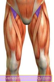
knee
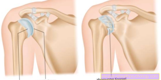
Osteonecrosis is also a typical disease of the knee, or the lower end of the thigh bone. If the knee is affected, one speaks of "Ahlbäck's disease" in medical terminology (Synonym: aseptic bone necrosis of the knee).
The cause of the death of the bone substance is primarily a disruption of the regular blood flow to the joint-forming thigh. This reduced blood flow is triggered by various mechanisms.
After serious accidents or violence, for example, tiny vessels can be damaged and the thighbone is undersupplied. In addition, it can be observed that osteonecrosis in the knee often occurs in patients with blood clotting disorders.
Regular consumption of tobacco products and / or alcohol can also be linked to the development of osteonecrosis in the knee. Although Ahlbäck's disease is a rather rare form of osteonecrosis, it could be concluded from observations of the disease cases that many patients had a history of treatment with bisphosphonates for osteoporosis. Accordingly, a connection between the long-term use of bisphosphonates and the occurrence of osteoporosis in the knee can be assumed.
The typical symptoms of Ahlbäck's disease are sudden, severe pain in the knee. This pain can arise after a minor injury or for no apparent reason. A patient who suffers from osteonecrosis of the knee does not only feel these pain phenomena during or after periods of stress. A typical symptom of Ahlbäck's disease is above all the so-called rest or night pain. In addition, physical exams in patients with osteonecrosis show swelling and limited range of motion in the knee. In such cases, treatment can usually be nonsurgical via hyperbaric oxygen therapy (in short: HBO), shock wave therapy and the use of pain relievers. Surgical replacement of the affected bone parts is only necessary in very pronounced cases.
Read more about this under Hyperbaric oxygen therapy
Ankle / talus
The ankle joint provides the articulated connection between the leg and the foot dar. Im upper ankle (Articulatio talocruralis) mainly articulate the lower (distal) ends of the Shin (Tibia) and des Fibula (Fibula) with the foot, especially with the so-called Ankle bone (Talus).
The most common form of osteonecrosis in the ankle or talus is the so-called Osteochondrosis dissecans. Osteochondrosis dissecans is a aseptic Bone necrosis one circumscribed Articular surface areas that can be associated with its rejection. So it comes to one near the cartilage Bone death. The disease occurs more frequently in the Growing age or in young adulthood and is associated with frequent injuries to the ankle (traumatic genesis) for example through "TwistYoung people also seem to be predisposed to that much sport float.
The osteochondrosis dissecans shows itself symptomatically by pain in the upper ankle joint, which is below burden occur and subside at rest. Therapy is usually conservative in children Immobilization possible, whereas in adults usually one operative re-fixation is inevitable. Therapy should be started as early as possible, otherwise it will wear out as the disease progresses cartilage and bone of the joint can come (arthrosis). In most cases, the ankle bone (talus) is affected by osteochondrosis dissecans. Most common is the top inner edge of the Talus (Trochlea tali, talus roll) affected. This bears most of the weight that is on the joint, which in turn indicates a load-dependent genesis of osteochondrosis dissecans.
Another special bone necrosis is the Renander's disease, in which the sesame bone of the big toe is affected.
jaw
The long-term use of bisphosphonates can lead to the death of bone tissue in all bony structures. While this phenomenon is quite rare in the knee area, bisphosphonate-induced osteonecrosis can be seen in the Jaws more often, however observe.
Furthermore, there are also drugs from the group of Steroids suspected of provoking osteonecrosis of the jaw and knee.
Patients suffering from osteonecrosis of the jaw usually have one marked instability of the bone on. Depending on the location of the dead bone, osteonecrosis can even cause Loss of perfectly healthy teeth to lead. In most cases, affected patients are treated by the Insertion of the body's own bone which is obtained from the alveolar ridge. If the osteonecrosis is less pronounced, this can also be done Immobilize help to stimulate the regeneration of the bone and restore the stability of the jaw. In children in particular, resting the jaw is the therapy of choice.
hip
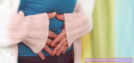
Also osteonecrosis of the hip is primarily through a Blood circulation disorder the bony structures triggered. In the area of Hip joint this circulatory disorder is mostly caused by high levels of fat in the blood, or a Disturbance of lipid metabolism provoked.
In addition, the regular one applies Consumption of tobacco products and or alcohol as a risk factor for the development of osteonecrosis of the hip.
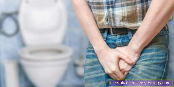
Affected patients usually complain at the beginning Pain in the groin. Classically take this Pain under exertion (while walking) increase significantly and decrease again during rest phases.
In order to avoid serious damage to the hip, if osteonecrosis of the hip is suspected, an comprehensive diagnostics be initiated. The therapy is divided into non-operational and surgical measures. First and foremost, the diseased hip should be treated with orthopedic aids relieved become. In addition, the intake appears Medicinal products that promote blood circulation in patients with osteonecrosis of the hip as useful. In severe cases, surgical treatment of the dead bone may be necessary.
Wrist (moon bone / scaphoid bone)
Osteonecrosis of the Moonbone (Os lunatum) will also Kienböck's disease or Lunate Malacia called. This is a aseptic Bone necrosis of the moonbone, which is located in the middle of the posterior (proximal) row of carpal bones. The lunar bone forms together with the other two proximal carpal bones, the Scaphoid (Os scaphoideum) and the Triangular leg (Os triquetum), and the Spoke (radius) of the forearm that wrist (Articulatio radiocarpalis). One is predisposing to osteonecrosis of the lunar bone mechanical load (e.g. using a jackhammer) or an anatomical variant in which the Ulna is shortened. The osteonecrosis manifests itself through Pain and restricted mobility. The doctor will also examine the affected bone Tenderness. Typical changes like Cysts, Contour changes or signs of osteoarthritis appear in the conventional roentgen only in later stages, which is why osteonecrosis of the lunar bone is suspected magnetic resonance imaging Imaging should be done. In the early stages the sufferer will be Painkiller prescribed and performed physical therapy. In later stages there is one operative joint stiffening (Arthrodesis) or the Shortening of the spoke (Radius) indexed. In rare cases, the moon bone must be surgically removed and replaced with silicone.
Just like the moon bone (os lunatum) can do that too Scaphoid (Os scaphoideum) be affected by osteonecrosis, one then speaks of one Preiser's disease. Those affected complain of stress-dependent pain with a projection onto the anatomical Snuffbox and the radial (thumb-facing) wrist region. Furthermore, it can lead to restricted mobility, overheating, swelling and reduced strength. Clinical tests are carried out for tenderness over the scaphoid bone. Here, too, depending on the stage, the therapy is conservative or surgical.
diagnosis

The diagnosis of suspected osteonecrosis begins with the Medical history survey (anamnese) and one physical exam. In this case, both the infected bone as well as the corresponding joint Function tests subjected.
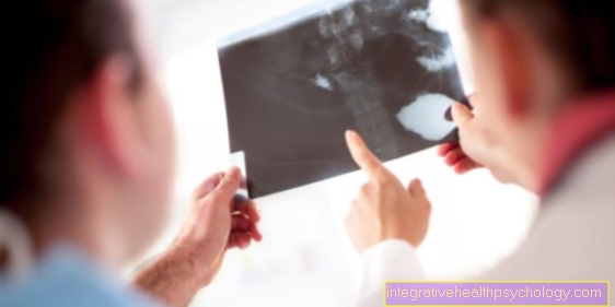
Then be imaging procedures used. Conventional methods like that Ultrasound examination (Sonography) and the X-rays usually only show typical changes in the bone in the later stages. "Osteonecrosis" can be diagnosed earlier and more reliably with the help of a Magnetic resonance imaging (Magnetic resonance imaging, MRI) which are best done with Contrast media is carried out.
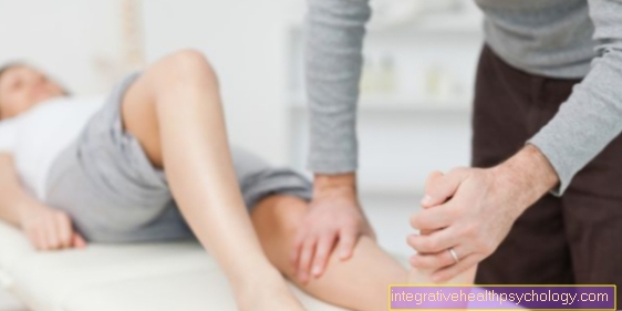
therapy
The therapy of choice for osteonecrosis depends on several factors. Sometimes it is enough for those affected To spare part of the body for a while and not to burden with weight, so to be treated purely conservatively.
Thanks to this break, it is not uncommon for one to be Spontaneous healing to reach.
In worse cases, however, only one can help surgical intervention. Again, there are several options. With small necroses, one can Drilling the bone (Pridie hole) suffice, otherwise also Bone graft (either with or without, depending on the infestation cartilage) and the use of Joint replacements (total endoprostheses, TEP) carried out.
course

The course of osteonecrosis differs from person to person. It is mainly dependent on:
- Age of the patient
- individually present Risk factors
- the trigger
- extent and localization of bone infarction
In general, the closer the dead bone tissue is to the joint and the larger this area, the more severe the course of this disease.
In principle everything is possible from the Spontaneous healing up to total destruction of bones.