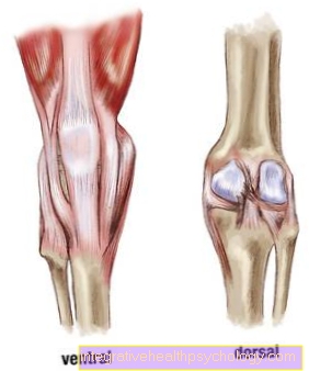
Osteonecrosis in the knee
introduction
Osteonecrosis is understood to be a Death of the bone. This is theoretically possible all over the body, but that knee belongs to the am most commonly affected joints. Women are affected somewhat more often than men (ratio around 3: 1).

causes
A Subdivision of osteonecrosis occurs in more septic and more aseptic Nature. An infection of the knee bone can occur, for example, after knee joint surgery. In aseptic bone necrosis, the death of the bone is not due to an infection with bacteria. The specific causes are not yet fully understood.
Circulatory disorders are considered a possible cause of aseptic bone necrosis. For example, in the event of repeated overloading or trauma due to minor injuries to the vessels, the bone can no longer be adequately supplied with oxygen from the blood. As a result, the insufficiently supplied tissue.
Another way of Reduced blood supply is a Embolism of the afferent arteries. This can be done, for example, as part of the Caisson diseasethat goes under the name Diving disease is known to occur. If a diver emerges too quickly, the artificially supplied breathing gases can clog arteries and thus lead to various long-term effects. One of these is that the bone dies over the weeks.
Risk factorsthat can promote the development of osteonecrosis are one radiotherapy of the affected knee in the past or a long-term cortisone therapy. Also be Alcohol consumption and Smoke discussed as a stimulus.
Appointment with ?

I would be happy to advise you!
Who am I?
My name is I am a specialist in orthopedics and the founder of .
Various television programs and print media report regularly about my work. On HR television you can see me every 6 weeks live on "Hallo Hessen".
But now enough is indicated ;-)
In order to be able to treat successfully in orthopedics, a thorough examination, diagnosis and a medical history are required.
In our very economic world in particular, there is too little time to thoroughly grasp the complex diseases of orthopedics and thus initiate targeted treatment.
I don't want to join the ranks of "quick knife pullers".
The aim of any treatment is treatment without surgery.
Which therapy achieves the best results in the long term can only be determined after looking at all of the information (Examination, X-ray, ultrasound, MRI, etc.) be assessed.
You will find me:
- - orthopedic surgeons
14
You can make an appointment here.
Unfortunately, it is currently only possible to make an appointment with private health insurers. I hope for your understanding!
For more information about myself, see - Orthopedists.
Symptoms
A typical symptom of knee osteonecrosis is Exercise-dependent movement pain. In severe cases, the knee can hurt even at rest. Additional symptom is a Knee joint effusion, which usually only occurs with larger defects and is caused by a swelling makes noticeable. Depending on the cause, for example an infection, you can also Signs of inflammation such as fever or overheating of the affected area.
Osteochondrosis dissecans
Osteochondrosis dissecans is one aseptic bone necrosis below the articular cartilage, which preferably affects the knee and in an unrestrained course in one rejection from Bone-cartilage fragments (Dissectate, articular mouse) the articular surface ends. If the articulated mouse becomes trapped, it may be responsible for a joint lock.
Ahlbaeck's disease
Ahlbaeck's disease is the name of the Osteonecrosis of the medial femoral condyle. The part of the knee joint formed by the thigh is the affected structure here.
He's going through diffuse pain, the usually gain weight quickly, noticeable. This does not result in the splintering of cartilage-bone fragments into the joint cavity as in osteochondrosis dissecans, but in one Collapse of the bone structure. The articular surface is deformed and the knee becomes one Varus deformity (If a) pushed.
Osteonecrosis of the knee in the child
It is with the child Specialty before that it is still growing is located. They are found above and below the knee cartilaginous epiphyseal platesfrom which new bone emerges and thus allows the bone to grow.
The disadvantage of Growth zone is that the Resilience not so great like a fully formed bone. For this reason, it is easier for children to become overburdened, especially when children do intense competitive sports. Correct ossification is therefore no longer guaranteed and osteonecrosis can occur.
Due to their growth potential, children have the advantage that the osteonecrosis can heal by itself if the affected knee joint is spared for a while.
Sinding-Larsen's disease
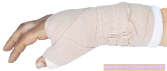
Sinding-Larsen's disease is an osteonecrosis that prefers primary school age boys concerns. The location of the necrosis is at lower end of the kneecap and hurts when exercised. There is no detachment of the lower end of the kneecap. In most cases, a break in sport is enough to allow the bone to heal the defect on its own, sometimes a splint can be worn to support the knee, which immobilizes the extended knee.
Osgood-Schlatter disease
Osgood-Schlatter's disease is one Osteonecrosis of the tibial headwhich often affects adolescent patients. It is believed that this disease is a Imbalance between the Resilience of the cartilage in growth and one excessive stress caused by sport.
In some cases, small bone fragments that can be found in the area of the quadriceps tendon become detached.
In this case, too, conservative treatment with relief until complete immobilization with a splint and crutches is sufficient. If small pieces of bone have loosened, they can be removed in a small surgical procedure after growth has ended.
diagnosis
The diagnostic starts with the physical examination. In addition to tenderness over the affected area, joint effusions or swellings can sometimes be noticed. If a joint mouse (Dissecat, detached fragment) becomes jammed, movement of the knee is painfully restricted.
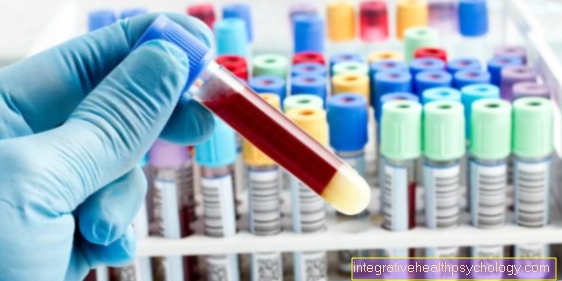
For the initial imaging, a X-ray image prepared. In the early stages this is usually without any findings. Only later, for example, will interruptions in the bony contour, newly formed bones in the form of light-colored sclerosis or the presence of a dissection become visible.
in the Early stage can have defects with one MRI getting discovered. When going to an investigation Contrast media enters the vascular system, a blood flow deficit in the area of osteonecrosis can be made visible. Small foci of the dissolving bone structure or fluid collections are also indicative.
In rare cases, a Scintigraphy performed with radioactive substances. At the beginning you see so-called cold lesions, in which the activity of the lesion is reduced compared to the surrounding bone tissue. In the further course, the defects are shown as more active as the new bone structure begins.
- MRI of the knee
- MRI with contrast agent - is it dangerous?
- Scintigraphy
therapy
Therapy depends on the stage and cause of the disease. To conservative therapy an observation is sufficient temporary immobilization and relief of the affected knee. In children and in the early stages, this is the best option.
Partly be Shock wave therapies and hyperbaric oxygen therapy used in patients to achieve a better regenerative capacity of the bone. So far there are no scientific studies that would prove a clear benefit.
If there is a septic bone necrosis the bacterium must immediately Antibiotics be treated. Since it is often difficult for this to get into the bone via the bloodstream, antibiotic chains, which release their active substance over a long period of time, are often and especially after an infection as part of an operation. In addition, foreign bodies such as a total knee endoprosthesis must be removed in case of doubt until the knee is sterile.
To operative therapy of knee osteonecrosis numerous possibilities. The primary goal is to preserve the joints. To revitalize the bones one can Drilling be made. Thereby stem cells immigratethat are supposed to fill in the defect.
Cartilage cell transplants can also be used for cartilage lesions. To fill in a bone defect, a Cancellous plastic respectively. For this purpose, bone is usually removed from the inside of the thigh and inserted into the necrosis zone. In the case of misalignments, surgery is usually performed to correct the joint axis in order to reduce the risk of osteoarthritis.
If there are extensive defects that cannot be treated with the above-mentioned procedures, the implantation of a prosthesis (e.g. knee TEP) is necessary.
forecast
At Children is the Very good prognosis. Due to their growth potential, the osteonecrosis heal very well if you take care of yourself.
With aseptic bone necrosis, the prognosis depends very much on the stage of the disease. In the early stages, the knee is fully functional after adequate therapy.
In later stages or difficult courses is the prognosis rather bad to see. The joint can usually only be replaced with a prosthesis during the course of treatment.
With septic bone necrosis, the prognosis is very different and worse than with aseptic necrosis, depending on how well the germ can be eliminated from the bone. In principle, however, the knee can again function fully.
prophylaxis
Since the ultimate cause of osteonecrosis on the knee not clarified is, no clear prophylaxis can be developed. Risk factors such as overwork and excessive, chronic alcohol abuse should be avoided if possible. With long-term cortisone intake, calcium and vitamin D preparations are also prescribed to prevent bone defects.