Quadriceps
Synonyms
Latin: M.. quadriceps femoris
German: quadriceps thigh muscle, four-sided thigh extensor, thigh extensor
introduction

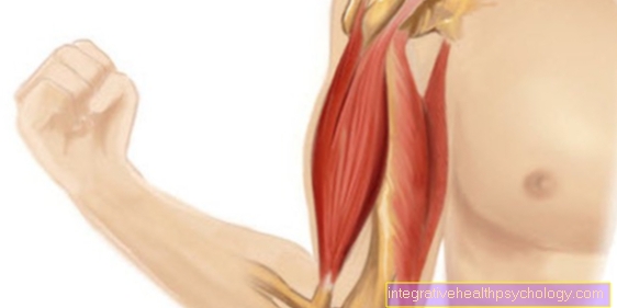
The quadriceps are the largest and most powerful muscle in our body Quadriceps femoris muscle is, as the name suggests, a muscle made up of four other muscles.
The physiological cross-section is more than 180 cm2 and weighs approx. 2 kg. It is divided into four independently emerging muscles. All four muscles of the quadriceps femoris muscle are located on the so-called ventral side or the front of the thigh. It is made up of:
- Even portion (Rectus femoris muscle) marked in blue
- Inner thigh muscle (Vastus medialis muscle) marked in green
- Outer thigh muscle (Vastus lateralis muscle) marked in yellow
- Middle thigh muscle (Vastus intermedialis muscle)
The straight part of the quadriceps shows significantly more almost twitch fibers in its physiological cross-section than the other three heads. It is therefore responsible for fast, vigorous movements, but is more prone to muscle fiber tears.
Rectus femoris muscle, M. vastus medialis, Vastus lateralis muscle and the M. vastus intermedius. With a width of approx. 150 cm2, it is one of the strongest muscles in the human body.
Rectus femoris muscle: It arises from the so-called Anterior inferior iliac spine or the acetabular roof of the hip joint and has its attachment to the anterior bone process of the tibia (Tibial tuberosity).
Vastus medialis muscle: Has its origin in the so-called Labium mediale the Linea aspera a line on the back of the thigh bone and also starts at the named bone process.
Vastus lateralis muscle: The origin of this muscle is this Labium laterale the Linea aspera and has the same approach as the other two muscles.
Vastus intermedius muscle: Its origin is at the front of the shinbone (tibia). Attachment also to the aforementioned bone process.
The nerve that "mediates" or innervates this function is the Femoral nerve. This is a peripheral nerve from the pelvic nerve plexus (Lumbar plexus) and arises from the spinal cord segments L1-L4.
Illustration quadriceps femoris muscle

Quadriceps femoris muscle
Four-headed hamstring muscle
(Quadriceps) - 1st + 2nd + 3rd + 4th
- Outer thigh muscle -
Vastus lateralis muscle - Upper thigh muscle -
Rectus femoris muscle - Inner thigh muscle -
Vastus medialis muscle - Middle thigh muscle -
Vastus intermedius muscle - Kneecap - patella
- Fibula - Fibula
- Shin - Tibia
- Femur - Femur
- Small rolling hill -
Lesser trochanter - Great Rolling Hill -
Greater trochanter - Iliac scoop - Ala ossis ilii
- Acetabulum - Acetabulum
You can find an overview of all Dr-Gumpert images at: medical illustrations

Thigh muscles
- Thigh Tie Tensioner -
Muscle tensor fasciae latae - Iliac muscle -
Iliacus muscle - Lumbar muscle -
Psoas major muscle - Comb muscle - M. pectineus
- Lean Muscle - M. gracilis
- Tailor Muscle - M. sartorius
- Upper thigh muscle -
Rectus femoris muscle - Outer thigh muscle -
Vastus lateralis muscle - Inner thigh muscle -
Vastus medialis muscle - Iliac-tibial tendon -
Iliotibial band - Kneecap - patella
- Long Dresser -
Adductor longus muscle - Big Dresser -
Adductor magnus muscle - Biceps thigh muscle,
long head -
Biceps femoris muscle,
Caput longum - Biceps thigh muscle,
short head -
Biceps femoris muscle,
Caput breve - Half-tendon muscle -
Semitendinosus muscle - Semi-membranous muscle -
Semimembranosus muscle - Femur -
Femur - Gluteus Muscle -
Gluteus maximus muscle
You can find an overview of all Dr-Gumpert images at: medical illustrations
Approach, origin, innervation
Approach: Roughness of the anterior tibial edge (Tibial tuberosity)
Origin:
- Straight part: anterior lower iliac spine (anterior inferior iliac spine) and the upper edge of the acetabulum
- Inner thigh muscle: distal (distant from the body) Rough line connecting the end of the two rolling hills (Linea intertrochanterica) and medial lip of the bony groin of the posterior femoral surface (Labium mediale linea asperae)
- Outer thigh muscle: large roll hillock (greater trochanter) and lateral lip of the bony groin on the posterior surface of the thigh bone (Labium laterale linea aspera)
- Middle thigh muscle: lateral surface of the thigh bone
Innervation: Femoral nerve, L 2 - 4
function
The function of the Quadriceps exists in an open function chain in a Elongation of the lower leg in Knee joint. This movement occurs when standing up, walking, running, jumping, and climbing. When the functional chain is closed, the function consists of catching and braking the upper body in the flexed knee joint.
So the function of the muscle is different. in the hip joint causes the Rectus femoris muscle a Flexion, so a flexion of the hip joint. It also supports and strengthens the hip joint.
in the Knee joint however, all 4 Muscles a Extension, i.e. an extension of the knee joint.
The Quadriceps femoris muscle is the only one Extensor of the knee joint. Its importance to the Straightening of the whole body is enormous. It also ensures that the kneecap remains in its slide channel.
At uneven development of the four muscles of the Quadriceps femoris In some cases, for example, previous injuries or illnesses can lead to Dislocation of the kneecap come, which means that there is a joint injury and the kneecap is no longer in contact with its articular surface.
The Opponent (antagonist) of M quadriceps femoris is the M. biceps femoris.
How is the muscle trained / contracted?
The Quadriceps is one of the most trained muscles in the Leg muscle training. Due to its high physiological cross-section, corresponding adaptation phenomena are to be expected.
In advanced Fitness training, as well as in the area Bodybuilding, is the quadriceps preferably through the Squats trained.
Due to the high coordination requirements of the squats are suitable for beginners and Health sector the following exercises: Leg press and Leg extension.
There are huge numbers of squat exercises. The most effective way to do this exercise is to do a Barbell on shoulders positioned. However, one should rather prefer low weight and the Slowly increase the number of repetitions. In addition, it is essential to note that both the Movement towards the floor as well as up from the hips takes place and always the Body tension is maintained. Wrong training in squats can to Back pain or. Injuries to lead.
Another simple method is the so-called "Climber" (Stair climber). It is not necessary to have (expensive) training equipment for this. Alternately if you push yourself with one leg on an elevation (e.g. a box) back and forth and changes the supporting leg. At the same time, the gluteus large muscles (Gluteus maximus muscle) stressed or trained.
Of course is too training on the leg press a popular method as well as effective method. Opposite the leg press, however, is here the strain on the knee joints greater.
Here you will find an overview of all relevant topics in strength training.
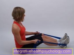
How is the muscle stretched?

The Thigh extensor stretching exercise is one of the most common exercises in sport. The Quadriceps femoris muscle is used in many sports (Soccer, to jog, tennis) heavily used. Therefore it requires an intensive stretching of this Muscle.
If the thigh muscles are not stretched sufficiently or correctly, this can lead to long-term Muscle shortening come. The strain is therefore essential if you want to avoid restrictions in locomotion.
The stretching ever 3 times run and for 20 seconds being held. It is recommended to do one between exercises Pause of approx. 30 seconds close.
It is a simple and very effective exercise Stretching of the muscle in Lateral position. The lower leg is turned 90 degrees in the Knee joint angled. The upper leg is held by the one on the same side hand at the foot and held the tension for 20 seconds. It is important to pull in the stomach and that pool to push forward until the stretching stimulus occurs.
Of course, this exercise can also be performed in the standing version, but this increases the likelihood that the pelvis will move.
Quadriceps tendon
The Quadriceps femoris muscle has many tendons that perform different functions.
The Tendon of the rectus femoris muscle starts about 10cm above the kneecap. This part of the muscle leads to Extension in the knee joint, as well as for Flexion in the hip joint.
The Tendon of the vastus intermedius muscle bundles together roughly in the middle of the thighbone (femur). Usually this is part of the muscle not visiblebecause it is completely covered by the rectus femoris muscle.
The Vastus lateralis tendon splits up and pulls on the side Kneecap over to the Lower leg.
The Vastus medialis tendon, which forms a common tendon of the four muscle parts, extends over the kneecap (patella) into the ligament of the patella, which attaches to a kind of roughening of the lower leg bone. The tendons themselves begin about 5-10cm above the patella.
Common quadriceps injuries
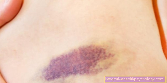
Injuries the front Thigh muscles in the context of stretching movements of the knee joint against resistance (e.g. a press blow during Soccer) can be used with pool- also with knee-joint injuries arise. Above all, play here Tears or tears (so-called ruptures) of the quadriceps tendon, the approaches of the Rectus femoris muscle and des Vastus medialis muscle as well as the Vastus lateralis muscle play an enormous role and lead to one decreased or to one Loss of extension of the knee joint.
Another cause of injury to the tendon of Quadriceps femoris muscle can degenerative be, i.e. that the tendon is in advance in its structure but also in hers Function damaged or. limited has been. This in turn can be achieved through the Tissue death (necrosis) or by perceptible Tissue shrinkage (atrophy) be conditional.
At intense tension of Quadriceps femoris muscle can it to further serious injuries come. In the Patellar fracture it comes to one Fracture of the kneecap. Despite this fracture, this can happen in individual cases knee can still be stretched, since this is where the tendon fibers of the Vastus lateralis muscle Start past the kneecap directly on the shin and allow for an extension (reserve extension apparatus).
In the other cases there is only one Injury to the articular surface of the kneecap before, in which the extensibility of the knee is hardly affected.