Striated muscles
Definition of striated muscles
Striated musculature is a certain type of muscle tissue because under polarizing light (e.g. a simple light microscope) it looks as if the individual muscle fiber cells are regularly striated.
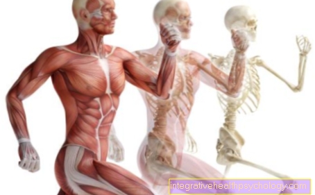
Usually the term is used synonymously for the skeletal muscles, as this type of tissue occurs primarily here.
Some muscles that are not responsible for moving the skeleton, such as the muscles of the diaphragm, tongue or larynx, are also of this type of tissue.
However, this transverse striation can also be found in the heart muscle, which, however, has some properties that are specific to it as well as some features that are not found in the rest of the striated muscles, which is why one usually speaks of three different muscle tissues: striated muscles, smooth muscles and heart muscle.

Types
There are two different types of striated muscles: the red and white muscles.
The muscle fiber cells of the red muscles have one high content of the oxygen supplier myoglobinwhich, thanks to its red color, is responsible for the color of this type of muscle. This is what makes red muscles above all for long-term loads designed and you can especially increase them at Endurance athletes how to find marathon runners.
Contain the muscle fibers of the white muscles however less myoglobin and therefore appear brighter. They are especially for fast, strong movements responsible and therefore predominate in people for whom muscle strength is particularly important, for example strength athletes.
Through training, white muscles can be converted into red, but whether this is also possible the other way round has not yet been conclusively clarified.
Structure of the striated muscles

Every skeletal muscle is from connective tissue (Epimysium) surroundfrom which individual fibers, which are also known as septa (partition walls), originate, which on the one hand surround each individual muscle fiber (Endomysium) and, on the other hand, group several muscle fibers together (Perimysium), so that the so-called Muscle fiber bundles form.
The epimysium passes into the muscle fascia and then into the Tendonsthrough which the skeletal muscle can be attached to the skeleton.
A distinction is made in the anatomy Insertion and origin of a skeletal muscle.
The horizontal striation is due to the special structure of the individual muscle fiber cells (myocytes). Apart from the usual cell organelles that can also be found in the muscle fibers (cell nucleus, mitochondria, ribosomes, endoplasmic reticulum (which here, however, is formed from a complex tubule system and is called sarcoplasmic reticulum)), these cells consist of thousands of so-called Myofibrils. These fibrils are thread-like structures that are tightly packed next to each other and run through the entire muscle lengthways. These are in turn made up of several sarcomeres.
Illustration of a muscle fiber

- Muscle fiber
of a skeletal muscle
Muscle fibra - Muscle fiber bundles -
Muscular Fasciculus - Epimysium (light blue) -
Connective tissue sheaths around groups
of muscle fiber bundles - Perimysium (yellow) -
Connective tissue sheaths
around muscle fiber bundles - Endomysium (green) -
Connective tissue between muscle fibers - Myofibrils (= muscle fibrils)
- Sarcomere (myofibril segment)
- Myosin threads
- Actin threads
- artery
- vein
- Muscle fascia
(= Muscle skin) - Fascia - Transition of muscle fibers
in tendon fibers -
Junctio myotendinea - Skeletal muscle
- Tendon fibers -
Fibrae tendineae
You can find an overview of all Dr-Gumpert images at: medical illustrations
Sarcomeres and contraction
Sarcomeres are a unit of the fibril which in turn consists of the smaller components Actin and Myosin consists.
Actin and myosin are Proteins, sometimes referred to as contractile proteins, as they ultimately allow our muscles to contract.
Actin and myosin are so regularly arranged in the sarcomeres that one certain pattern arises:
Both actin (directly) and myosin (via another, very flexible protein) are attached to the so-called Z-washers.
From these disks there is an area called "Iban de“, Which usually only contains actin. This area therefore appears brighter under the light microscope than the "A bands". These represent the area where actin and myosin overlap, more or less depending on the state of contraction of the muscle.
If the muscle is relaxed, you will find a place that "H zone“, Which only has myosin but no actin. When the muscle is contracted, however, the myosin filaments move closer towards the Z-disks, which is why they then overlap more and more with the actin filaments and the "H-zone" becomes shorter and shorter until it finally disappears.
This process is called in medicine Sliding filament mechanism known and is the Basis for our muscles to shorten.
For this process to take place, the muscle needs on the one hand Calcium ions, which he receives on the one hand from the sarcoplasmic reticulum and on the other hand from the cell environment, and on the other hand the Energy supplier ATP.
If no more ATP is formed, the contraction of the muscle can no longer be released, which is why it remains in this tense state. This happens when an organism dies and the body remains in a rigor mortis.
Excitation of the striated muscles
An important feature of the striated muscles, precisely in order to differentiate them from the smooth muscles and the heart muscles, is that they are ours arbitrary control subject. ´
Striated muscles can get from us consciously tense or relaxed become.
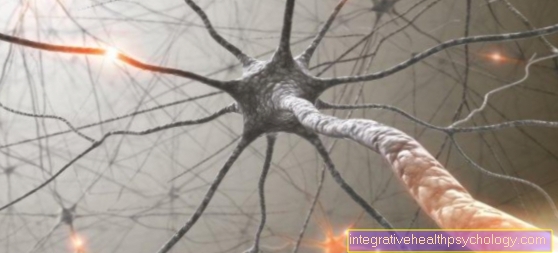
You will be from motor nerve fibers reached, at the end of which a neuromuscular endplate lies. Here is the distribution of a Carrier substance (Transmitters) named Acetylcholine. This binds to receptors that are located on the muscle, which ultimately leads to channels opening there that lead to a discharge of the muscle cell:
A so-called is formed Action potential which is passed on via the membrane of the muscle cell, through which calcium finally reaches the interior of the cell in several steps, where it sets the sliding filament mechanism in motion. The muscle contracts.
In most muscles, the individual muscle fiber cells are excited by a large number of nerve cells. Depending on how many nerve cells are activated, different numbers of muscle fibers contract within a skeletal muscle, not always the entire muscle. Furthermore, the body is able to do that control the muscle power required at the moment. Just because you want a muscle to be active doesn't mean it needs full strength.


