Thoracic spine pain
introduction
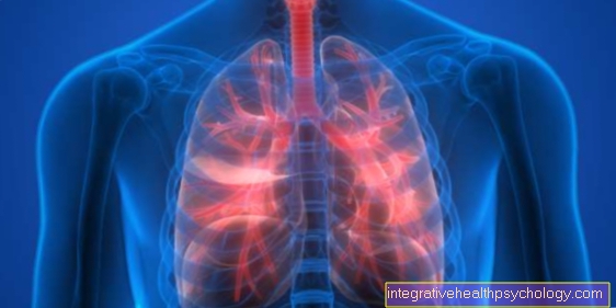
The thoracic spine consists of 12 vertebrae and is located between the cervical and lumbar spine. Symptoms in the thoracic spine area are usually described by those affected as dull or pressing pain, especially between the shoulder blades. Due to the articulated connection of the vertebrae in the chest area and the ribs, the pain can be movement-dependent depending on the cause. The pain emanating from the thoracic spine can also radiate into the chest in a belt-shaped manner.

General causes
Compared with other sections of the spine, the thoracic spine causes it fewer complaints. Through the stable rib-vertebral joints and participation in the bony rib cage, the thoracic spine is in hers Relatively limited range of motion. As a result, e.g. the risk for Herniated discs of the thoracic spine to below 2%. However, there are diseases that directly affect the thoracic spine or include it. In addition, sometimes also neighboring organs, such as. the heartCause pain in the thoracic spine area.
Appointment with a back specialist?

I would be happy to advise you!
Who am I?
My name is I am a specialist in orthopedics and the founder of .
Various television programs and print media report regularly about my work. On HR television you can see me every 6 weeks live on "Hallo Hessen".
But now enough is indicated ;-)
The spine is difficult to treat. On the one hand it is exposed to high mechanical loads, on the other hand it has great mobility.
The treatment of the spine (e.g. herniated disc, facet syndrome, foramen stenosis, etc.) therefore requires a lot of experience.
I focus on a wide variety of diseases of the spine.
The aim of any treatment is treatment without surgery.
Which therapy achieves the best results in the long term can only be determined after looking at all of the information (Examination, X-ray, ultrasound, MRI, etc.) be assessed.
You can find me in:
- - your orthopedic surgeon
14
Directly to the online appointment arrangement
Unfortunately, it is currently only possible to make an appointment with private health insurers. I hope for your understanding!
Further information about myself can be found at
diagnosis
So that the attending physician can reliably assign the pain to the thoracic spine, he must use various examinations and methods.
1. Medical history
At the beginning of each examination, a detailed medical history (Greek anamnesis = memory) respectively.
For this purpose, the patient is asked in detail about his complaints.
In particular, the thoracic spine will be accurate Localization of pain, (e.g. Vertebral body height, lateral, central, belt-shaped), Pain quality (dull, stabbing, burning, pulling etc.), Pain occurring (e.g. breath-dependent, movement-dependent, spontaneous, pressure-sensitive), Duration of pain (Hours, days, weeks etc.), as well as any Accompanying complaints such as. neurological or other abnormalities (Numbness of the arms, paralysis, incontinence, fever) he asks.
So many conclusions can already be drawn about the causes of the pain in the thoracic spine!
2nd inspection
In the second step, the doctor examines the entire spine on the undressed upper body. He pays particular attention to this symmetry and visible external changes or Injuries.
E.g. Oblique shoulder position, Reference to a Scoliosis be in the area of the ESPE. On the other hand, if there are small, red blisters in the painful area, it is probably one Shingles.
3. Manual examination
This generic term includes examinations of mobility or else Pain provocation tests.
First of all, the doctor can gradually palpate the thoracic spine to check whether there is a knocking sensation. Pressure sensitivity (as with blockages or vertebral inflammation) is available.
The condition of the possibly hardened muscles can be detected by palpation (palpate) capture.
To assess mobility, the orthopedist often asks e.g. to rotate or bend the thoracic spine. The painfulness of these exercises provide further valuable information!
To check the neurological status (e.g. limited at Herniated disc of the thoracic spine with involvement of the nerves) a strength test of the arms can be carried out.
4. Imaging procedures
In the last place in the diagnosis of thoracic spine pain are imaging tests.
They are usually indicated if the pain has lasted for more than a week or if serious complications (e.g. Paralysis of the arms) occur.
Depending on the question, X-ray examinations, MRI images of the thoracic spine, CT-Recordings, Myelographs or Scintigraphy are used.
5. Further procedures
If there is a suspicion of an inflammatory or tumorous event, you can Blood tests be performed.
In individual cases, a thoracic vertebra or intervertebral disc puncture can be prescribed. In addition, it should be clarified whether there is a connection between the back pain and internal organs. Sometimes you can Heart attacks or Pneumonia Cause discomfort in the thoracic spine.
Therapy measures in general

The basic requirement for a successful therapy is the exact one Root cause research. Because only when the pain-causing event has been clearly identified, a targeted and individual treatment can take place.
1. Pain Management
When those affected are acutely affected by pain in the thoracic spine, this occurs in the vast majority of cases pain reliever drugs for use.
Otherwise there is often a "Vicious circle“.
Due to the massive complaints, we take, often unconsciously, a supposedly more bearable one Relieving posture a. As a result of this unnatural posture, the already tense muscles cramp and cause even more more pain!
As a rule, treatment begins with the group of "non-steroidal anti-inflammatory drugs", short NSAIDs. They work both pain reliever, as well as anti-inflammatory on the affected thoracic spine section.
The known active ingredients include e.g. Ibuprofen or Diclofenac.
However, caution is advised with prolonged administration!
Taken over the long term, they can cause addiction and numerous undesirable side effects. So the NSAIDs also inhibit the formation of the protective Mucus layer of the stomach.
If taken for months, the aggressive stomach acid begins to attack the surrounding walls. In the worst case, arise chronic inflammation of the gastric mucosa (lat. gastritis) or Stomach ulcers (Latin ulcer)!
If you take painkillers on a long-term basis, you can the Kidneys and liver be harmed.
Basically, it is important to keep the duration of intake as short as possible! Alternatively to the prescription of tablets, pain reliever or numbing can be used Anoint are used.
They have the great advantage of not causing any serious side effects, as they only work locally.
If patients suffer from long-term pain in the thoracic spine, pain pills are unsuccessful, or there is a risk of long-term use local narcotics represent an alternative. To do this, the doctor injects pain-relieving medication under the skin or into the muscles with a fine needle. Injections are particularly promising if the pain is precisely localized (Trigger points).
Stubborn and therapy-resistant complaints (e.g. Intercostal neuralagias) can be alleviated by deeper injections directly into the bone or joint (thoracic facet infiltration, spinal nerve analgesia, costotransversal blockade).
It is hoped that this will result in a direct elimination of the responsible pain sensors or nerve roots.
The procedure takes place with full consciousness, only the puncture site is anesthetized. Most of the time, the injection is carried out with the patient sitting and the back slightly arched forward.
At all costs one should current x-ray image present! Such procedures must be carefully considered because of complications, such as infections or Cardiovascular problems can take place.
2. Heat application
Often there is pain in the thoracic spine, muscular tension underlying.
The application of heat stimulates the blood flow to the affected muscle areas and thus relieves the cramps.
Numerous variants of the so-called "Heat patch“Offered. Contact with the surface of the skin or oxygen from the ambient air stimulates chemical processes that generate a pleasantly perceived heat development of around 40 degrees.
3. Physiotherapy
In principle, physiotherapy is very useful in many cases! Because in addition to muscular tension, the pain is often caused by small blockages Rib or vertebral arch joints underlying.
The aim of the therapy is to release these blockages and relax the muscles. Because very often there is a close connection between the two phenomena. The physiotherapist can do this Massage techniques, Strengthening exercises or else Taping apply.
Overarching goals are mostly to recognize posture errors and incorrect movement patterns and then to correct them.
Ideally, the patient is given targeted guidance during physiotherapy so that he can also carry out the exercises independently in everyday life.
4. Operations
Particularly severe cases, such as Thoracic spine tumors, Vertebral body infections or severe scoliosis can make an operation essential.
As a rule, however, the benefits and risks of such an intervention should be carefully weighed against each other. In addition to undesirable accompanying complications, they often do not achieve the desired success!
Possible causes and targeted therapy
To the possible causes that can lead to thoracic spine pain include:
- Scoliosis
- Degeneration and blockages
- Intercostal neuralgia
- Spondylitis, spondylodiscitis
- disc prolapse
- Injuries to the thoracic spine
- Thoracic spine tumors
Scoliosis
When viewed from behind, the normal spine is straight. At a Scoliosis, however, lies a lateral bending or curvature in front. The incidence of the disease is stated very differently and fluctuates between 0.13% and 13.6%. What is certain is that Girls are about four times more likely to be affected than boys are.
In a majority of scolioses, the exact one is The cause is still unknown today (idiopathic). It is believed that during growth, especially during puberty, the Vertebral bodies grow unevenly and asymmetrically. This creates a Rotation or twist (Torsion) The spinewhich naturally shouldn't be there. Due to the initial painlessness in childhood, scoliosis often becomes accidental, e.g. during physical education or by parents. Due to the misalignment, the children often have one accompanying them shoulder-or Pelvic inclination. The severity of scoliosis can vary greatly: in the majority of cases, the spine is only slightly crooked and at most causes it cosmetic problems. On the other hand, a fully developed scoliosis, if left untreated, very severe deformities and cause health problems!
In principle, all sections of the spine can be affected. Most often, however, one finds displacements and deformations of the thoracic spine. The connection with the ribs creates a so-called "Rib hump". During the growth phase, those affected very rarely experience pain. However, due to the constant improper stress already arise in young adulthood painful signs of wear and tear. With age, the pain in the thoracic spine leads to a instinctive relieving posture. This will make the back muscles heavily overused and causes additional discomfort. The deformation can go so far that both breathing and cardiac output are restricted.
The decisive factor for the therapy of scoliosis is Severity of the bending. Slight curvatures can be targeted with physical therapy be treated. If you suffer from advanced scoliosis, you can wear one Corsets or one surgery be displayed.
Early detection is therefore of great importance! Because only if scoliosis is detected in time can pain in adulthood be prevented. The pediatrician examines children aged 9-10, e.g. through the "Prevention test", the correct curvature of the thoracic spine: To do this, the child bends as far forward as possible with bare upper body and closed, straight legs.Asymmetries or differences in level, such as a rib hump, can be recognized.
Degeneration and blockage
In the thoracic spine area, we find two different types of joints: The small vertebral arch joints (Articulatio zygapophysiales, Facet joint, vertebral joint) are located in pairs between the articular processes of two adjacent vertebrae. she guarantee great mobility within the spine. In the thoracic spine area, however, the range of motion is greatly reduced compared to the remaining sections. The cause of the restricted mobility are Rib head joints (Articulatio capitis costae), as well as the Rib cusp joints (Articulatio costotransversaria). They are each formed by a part of the rib and small joint surfaces of the thoracic vertebrae. In the totality arises in this way, together with Sternum, of the bony rib cage (thorax)
Find degenerative now, that is changes due to wear instead, they primarily affect the joints of the thoracic spine. Herniated discs (Herniated disc), as e.g. often occur within the lumbar spine, but are extremely rare. The small joints of the thoracic spine can therefore be affected with increasing age, with permanently incorrect stress or posture.
In this context one often speaks of "Joint blockages". As a result of the degenerative damage to the articular surfaces described, but also due to Changes in muscles and ligaments, can temporarily Blockages occur. In addition to restricted mobility, are Concomitant symptoms such as. Characteristic pain in the affected area. Often describe patients belt-like pain and pronounced pressure sensitivity in the affected area.

Side view motion segment
- Vertebral bodies
- Intervertebral disc
- Spinal nerve root
- Intervertebral hole (neuro foramen)
- Vertebral joint
- Spinous process of the vertebra (palpable on the back as the rear end of the vertebra)
Find degenerative now, that is changes due to wear instead, they primarily affect the joints of the thoracic spine. Herniated discs (Herniated disc), as e.g. often occur within the lumbar spine, but are extremely rare. The small joints of the thoracic spine can therefore be affected with increasing age, with permanently incorrect stress or posture.
In this context one often speaks of "Joint blockages". As a result of the degenerative damage to the articular surfaces described, but also due to Changes in muscles and ligaments, can temporarily Blockages occur. In addition to restricted mobility, are Concomitant symptoms such as. Characteristic pain in the affected area. Often describe patients belt-like pain and pronounced pressure sensitivity in the affected area.
In manual medicine and chiropractic therapy, such blockages are released through targeted mobilizations. Experienced Chiropractors, can often restore full mobility within a few minutes. However, it can take several days for the Muscle tension and pain subside. To Pain relief, sufferers therefore often take medication, such as Anti-inflammatory drugs (e.g. ibuprofen). However, if a major injury is suspected, such as If there are fractures in the thoracic spine area, manual therapy must not be used under any circumstances. Because the jerky and powerful movements of the therapist can worsen existing fractures (e.g. vertebral body fracture) under certain circumstances.
Since the Ribs connected to the thoracic spine local blockages can be extremely uncomfortable. Because through our breathing, the chest rises and falls permanently and provokes pain in the blocked costal vertebral joint. Not infrequently, the physician takes such painful conditions as Thoracic spine syndrome (short: BWS syndrome) together.
Intercostal neuralgia
At the lower edge of the ribs, the Intercostal nerves (Intercostal nerves), with the lowest 12th rib of the Subcostal nerve speaks. With the clinical picture of Intercostal neuralgia, sufferers feel belt-shaped pain radiating from the thoracic spine or anterior chest. Often they come Sudden and seizure discomfort. Not infrequently, additional ones occur Parasitic sensations or numbness in the affected area on.
Causal, are common wear-related changes in the thoracic spine to find. Typically, the pain can be relieved by certain positions, e.g. Rotational movements, are provoked. First and foremost, the therapy depends on the patient's level of suffering. By Pain medication, possibly too Injection therapy with local anesthetics, intercostal neuralgia can be treated.
In any case, the symptoms described must also include diseases that are not related to the thoracic spine. E.g. of the Herpes zoster virus ("Shingles") Similar symptoms, but accompanied by typical reddish and blistered rash. But also with acute ones Heart attacks, Pulmonary embolism or other organ diseases may initially hurt the thoracic spine.
Spondylitis, spondylodiscitis

If a Vertebral bodies inflamed, one speaks of a Spondylitis. Is also still the adjacent disc affected, it is a spondylodiscitis.
First and foremost, stand general complaintssuch as fever, night sweats, loss of appetite or fatigue in the foreground. In addition, there is an extreme Tension and tapping pain in the area of the affected vertebral body. Due to the massive infection in the body, the classic ones Inflammation values (BSG, CRP) increased in blood. There used to be a great risk that the spondylitis would not be recognized in time. Permanent symptoms of paralysis were not infrequently the result.
Fortunately, it is now possible to treat the disease early enough in almost all cases. So the prognosis is through strict bed rest, plaster casts, drug treatment and Operations Cheap.
Herniated disc of the thoracic spine
Herniated discs in the thoracic spine area are extremely rare and a real rarity. In addition, cases that occur are usually easy to treat. If a herniated disc still causes symptoms, those affected describe severe pain in the affected segment. In very rare cases, neurological complaints are added. These include discomfort, paralysis and sudden incontinence. As soon as such symptoms occur, in addition to the actual pain in the thoracic spine, you must immediately consult a doctor.
Read more on the topic: Symptoms of a herniated disc of the thoracic spine
Injuries to the thoracic spine
As Result of sports or leisure accidents can be simple Fractures of the vertebral bodies arise. Often it is injuries that result from serious traffic or work accidents.
After such events, they are almost always evident Hematomas ("Bruises") in the area of the broken bone. Affected people sometimes feel severe pain and pronounced tenderness.
Fortunately, there are mostly stable fractures in the thoracic spine, where no neurological complications to be expected are. In such cases it is sufficient for a certain period of time bed rest to hold on a flat surface and then physiotherapy exercises perform.
Thoracic spine tumors
The most common tumors in the thoracic spine are Metastases, one rarely finds Primary tumors. The underlying diseases can e.g. Tumors in the area of Count your thyroid, lungs, prostate, or chest.
Those affected usually go to the doctor because they have one dull pain feel in the affected part of the spine. Often there is a so-called "Shaking pain": Physical activities that involve small shocks in the spine, such as Jogging or jumping cause the pain described. Another clue can be sudden fractures in vertebral bodies. If the tumor spreads, it can also press on the adjacent spinal cord or the emerging nerve roots. Diverse neurological symptoms can result.
anatomy
The spine can be folded in four sections structure. In addition to the thoracic spine, the cervical and lumbar spine as well as the sacral spine (sacrum) form a functional unit.
In the middle area we find the thoracic spine. it consists of 12 vertebral bodies and together with the 12 Pairs of ribs and the sternum (sternum) the bony rib cage.
Naturally, the thoracic spine describes one convex curvature to the rear (dorsal), called kyphosis. In contrast, the cervical and lumbar spine are forward (ventral) convexly curved. The doctor then speaks of a lordosis.
Illustration of the thoracic spine

Thoracic spine (green)
- First cervical vertebra (carrier) -
Atlas - Second cervical vertebra (turner) -
Axis - Seventh cervical vertebra -
Vertebra prominent - First thoracic vertebra -
Vertebra thoracica I - Twelfth thoracic vertebra -
Vertebra thoracica XII - First lumbar vertebra -
Vertebra lumbalis I - Fifth lumbar vertebra -
Vertebra lumbalis V - Lumbar cruciate ligament kink -
Promontory - Sacrum - Sacrum
- Tailbone - Os coccygis
You can find an overview of all Dr-Gumpert images at: medical illustrations

.jpg)