Swinging Flashlight Test
introduction
The Swiniging Flashlight Test is a very easy-to-use test from neurology and ophthalmology. It is used to detect what is known as a relative afferent pupil deficit (RAPD), which can occur when there is inflammation behind the eye - retrobulbar neuritis. Typical diseases in which RAPD occurs are multiple sclerosis (MS) and neuromyelitis optica (NMO-SD).

definition
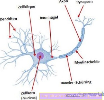
The Swinging Flashlight Test is a medical diagnostic procedure that is very easy to carry out and which only requires a small flashlight (ideally a pupil light). The test is done on patients in ophthalmology or neurology to detect a disorder of the optic nerve. For the sensitive perception and processing of sensory impressions of the eye, the information is passed through so-called "afferent nerve fibers" to the brain in order to trigger reflexes there or to forward the information to the cerebral cortex to become aware of what has been seen. Opposite them are the so-called “efferent” fibers, which direct a corresponding reaction from the brain to the rest of the body, in this case the eye, to bring about a change in the size of the pupil, for example. The Swinging Flashlight Test detects disorders of the afferent nerve fibers, which can occur, for example, with inflammation of the optic nerve (retrobulbar neuritis) in MS or with brain or eye injuries.
Find out more about the Structure of the nervous system
Indications
The Swinging Flashlight Test can quickly determine whether there is a sensitive perception disorder in an eye, which is referred to as a relative afferent pupil deficit (RAPD). This test can be necessary for various diseases of the eye, the visual tract or the brain. In the eye, diseases such as macular degeneration, cataracts or glaucoma, as well as retinal diseases can cause perception disorders in the eye. Furthermore, diseases of the visual tract, such as inflammation of the optic nerve or compression of the intersection of the visual tract, can cause a noticeable swinging flashlight test. Various neurological diseases can also restrict sensitive processing in the brain and result in a conspicuous test result. In acute accidents, the Swinging Flashlight Test can provide information on a traumatic brain injury with damage to the visual pathway.
preparation
The patient does not have to be awake to prepare for the swinging flashlight test; the pupillary reflex can usually be triggered even in an unconscious patient, if one disregards deeply comatose states. Only a pupil light or a small flashlight is necessary for this. In order to be able to carry out the test as precisely as possible, the room should be darkened so that the pupils are as wide as possible at the start of the test. In order to clarify an unclear result more precisely, gray filters can still be held in front of the eyes in order to make the differences in light more clear.
procedure
The execution of the swinging flashlight test is basically very easy and can be done within a few seconds. In the ideally darkened room, if the patient's pupils are wide, the lamp alternately shines into one and the other eye. It is swung from one eye to the other within 1-2 seconds, which in healthy patients causes the pupils to reflect light. With healthy, sensitive perception of the eye, the illumination of the eye causes the pupils to narrow on both sides (!) When light falls, in order to reduce the amount of light entering the eye. While the lamp is swung to the other side, i.e. for a brief moment without incidence of light, the pupils should widen again in order to narrow again when illuminated.
evaluation
In healthy people, the pupillary reflex is triggered on both sides when light falls into one of the two eyes. This means that when the left eye is illuminated, there is also a constriction of the pupil in the right eye, which is referred to as one consensual light reaction. If there is a disorder of the so-called “afferent nerve fibers”, there is neither a pupillary reflex in the same nor in the neighboring eye, since the information from the incident light no longer “penetrates” and can no longer trigger the pupillary reflex. In the Swinging Flashlight test, the quick change of the side causes both pupils to widen again and again if the light no longer falls briefly into one of the eyes. Normally, both (!) Pupils should contract again as soon as the light hits a pupil again. In the event of a defective sensory perception of the visual path on the illuminated side, the pupils remain dilated, since the information from the incident light no longer “penetrates” and can no longer trigger the pupil reflex. If, on the other hand, the healthy eye is illuminated again, the pupils continue to contract.
What are the alternatives?
The Swinging Flashlight Test is a test that can be carried out quickly, but it has only a relatively low informative value. In addition, it is often not easy to assess, especially with mild findings, and requires a certain amount of experience.
The exact illumination of both eyes with renewed darkening of the eyes in between, however, in principle enables a differentiation between a so-called “afferent” and “efferent” visual path disorder. Patients with retrobulbar neuritis (RBN) in the context of an MS attack also usually report unilateral visual disturbances.
To further investigate the visual pathway, the visually evoked potentials should be measured. For this purpose, changing light stimuli are offered to the patient, which then lead to a measurable reaction in the visual cortex of the brain. If this reaction is slowed down, an afferent visual path disorder can be assumed. In addition, if a disorder is present, an imaging procedure such as, ideally, a skull MRI must be performed for precise localization.
Further information
- Structure of the nervous system
- Inflammation of the optic nerve
- multiple sclerosis
- Skull MRI


