How can you recognize a pulmonary embolism? What are the typical signs?
introduction
Pulmonary embolism can be associated with typical symptoms. However, these do not necessarily have to be available. If a pulmonary embolism is suspected, a doctor should be consulted immediately. A physical exam and imaging tests can help doctors determine if the lungs have had an embolism.

Physical symptoms
A pulmonary embolism presents itself as a variety of physical symptoms, which, however, do not have to be identical in every person. Accordingly, a pulmonary embolism cannot always be clearly diagnosed clinically without further diagnostic measures being taken. It is particularly important to see a doctor when there are symptoms of a leg vein thrombosis so that a pulmonary embolism cannot even occur due to detached thrombus parts.
A pulmonary embolism itself presents itself with different symptoms depending on the severity. In the mildest stage, those affected usually feel no symptoms at all. If it is more severe, the typical symptoms of acute shortness of breath and chest pain, which can also occur depending on the breath, can occur. The pain is then particularly evident when inhaling. Other clinical symptoms include a rapid pulse (Tachycardia over 100 beats per minute), coughing with possibly bloody sputum, a high breathing rate (Tachypnea), Cardiac arrhythmias and low blood pressure.
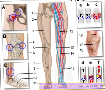
These symptoms can vary in severity and can even lead to shock. However, they do not necessarily all have to be present. In order to recognize a pulmonary embolism, the observation of the physical symptoms is crucial. In doing so, it is essential to watch out for signs of a thrombosis in the legs, which can be expressed, for example, by reddening, swelling, overheating and / or tenderness in a leg.
Read more on the topic: Symptoms of pulmonary embolism
Back pain
Back pain is not necessarily a typical symptom of pulmonary embolism, but if there is sudden severe pain in the thoracic spine, it should also be clarified whether a pulmonary embolism is present.
Usually, pulmonary embolism is accompanied by chest pain. These can vary depending on the size of the embolism. Recurring pain can occur as a harbinger of pulmonary embolism. In acute cases, sharp pain usually occurs suddenly. Since the nerve fibers from the back and chest area have a common connection point, pain originating in the chest can also be mistakenly perceived in the back.
to cough
Cough is one of the many symptoms that can occur with a pulmonary embolism. Often times, the cough is associated with rattling breath. A cough with bloody sputum is also a possible symptom. A cough can also be a sign of the signs of a pulmonary embolism.
The pulmonary embolism usually comes from a blood clot that lodges in a lung vessel. However, before this entire clot dissolves and reaches the lungs, smaller parts of the clot can be washed into the pulmonary vessels. There they are not immediately noticeable with the typical symptoms of a pulmonary embolism, but symptoms such as coughing and wheezing do occur.
Diagnostic measures to detect pulmonary embolism
Eye diagnosis / suspicion
In order to recognize pulmonary embolism at an early stage, it is important to know the symptoms and learn to perceive them in yourself.
Only patients who know what a pulmonary embolism might present can see a doctor in good time if it shows early signs. The physical symptoms are therefore in the foreground in early detection. If you experience sudden breathlessness, which may be accompanied by chest pain and swelling of one leg, you should go to a hospital as soon as possible.
Further diagnostics can then take place there. Visiting a hospital in good time is crucial for the success of the therapy. Even if a thrombosis is suspected, a doctor should be consulted at an early stage so that the blood clot in the leg can be professionally treated. In this way the development of a pulmonary embolism can be prevented.
More about this topic can be found: Causes of pulmonary embolism
Computed Tomography
If a pulmonary embolism is suspected, a Computed Tomography (CT) can be made. It is the fastest way to secure the diagnosis. For this purpose, the patient is pushed into a kind of tube that is made of X-ray cross-sectional recordings of the body. In order to make the blood clot in the pulmonary vessels particularly clearly visible, the patient is given a Contrast media injected. The extent and location of the pulmonary embolism can then be determined on the images.
Computed tomography may miss small embolisms. However, you can use Lung scintigraphy be made visible.
EKG changes
If a pulmonary embolism is suspected, an ECG is usually written early.
For this purpose, various electrodes are attached to the patient's chest in a specific arrangement. The electrodes conduct the electrical currents above the heart. These are recorded on paper in the form of a curve, which gives the doctor indications of the condition of the conduction of excitation in the heart muscle. In the case of a pulmonary embolism, there are typical signs on the EKG that indicate this disease. One speaks of the so-called SIQIII type. The name refers to a special form of the EKG curve that has S-waves in the first lead and Q-waves in the third lead. In addition, the ECG often shows a rapid heartbeat in the case of a pulmonary embolism (Tachycardia) and cardiac arrhythmias.
Read more about our topic: EKG changes in a pulmonary embolism
Can a pulmonary embolism be recognized on an X-ray?
A conventional chest X-ray is a rather minor method for diagnosing pulmonary embolism, since a CT image of the chest can usually provide much more specific information.
Sometimes a chest X-ray may initially be made in order to rule out other causes of the symptoms. However, if the suspicion of pulmonary embolism is confirmed, a CT chest is usually also performed.
Read more on the topic: Chest x-ray (chest x-ray)
Signs of a pulmonary embolism, which could possibly be seen on a chest x-ray, are, on the one hand, a pleural effusion as a sign of increased vascular permeability in the event of a congestion of the blood, an enlargement of the heart shadow as a result of increased stress on the right heart, and various signs of a pulmonary infarction if it is caused by the reduced blood flow lung tissue death has already occurred.
Under certain circumstances, however, these changes can also be present in the context of other diseases, so that the final detection of a clot in a pulmonary embolism is usually only possible with great certainty within the framework of a CT angiography.
Further diagnostics
To further confirm the diagnosis of pulmonary embolism, a Blood count be made. This is based on the so-called D dimers examined. These are fission products of fibrinwhich is found in blood clots. If the body is busy breaking down such a thrombus, as in the case of a pulmonary embolism, the D-dimers in the blood are increased. With normal D-dimers in the blood, pulmonary embolism can be ruled out with great certainty.
Furthermore, a Magnetic resonance imaging (MRI) or a Angiography pulmonary circulation to diagnose pulmonary embolism.


.jpg)