Central venous catheter
definition
A central venous catheter, or CVC for short, is a thin tube that is pushed through a large vein to just before the heart.
The other end is free outside the body and usually consists of several accesses. These can be used to administer fluids (infusions) and medication and to take blood. In addition, the pressure in the venous system can even be measured.
A CVC is placed in the hospital, for example during major operations.
As bacteria could enter the body when the catheter is inserted, careful hygiene must be observed and the catheter removed if there is a fever or other signs of infection.

indication
A central venous catheter is usually placed when a safe and large access to the circulatory system is required. The reasons for this are diverse. For large and long-lasting operations,
which may subsequently require care in an intensive care unit, a CVC is often placed in advance.
An indication can also arise if another access route via a needle in the arm is not possible due to poor vein conditions. There are also liquids (infusions),
which irritate the smaller veins, so they should be administered through a large central vein using a central venous catheter.
Artificial liquid nutrition via the blood should also be supplied via a central access. There are also drugs that are supposed to work directly on the heart and get as close as possible to it. A ZVK is also indicated for this.
In addition to this use of the catheter as an access route, there are other possible uses and thus indications. With the help of special measuring devices, the
Measure central venous pressure and oxygen saturation. This allows a detailed monitoring of important body function values, for example of patients who are being treated in an intensive care unit.
Puncture sites
For the placement of a central venous catheter, there are basically various points on the body available and the doctor can choose the most suitable for the respective patient.
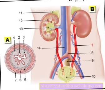
The prerequisite for choosing a vein is that it is large enough and that the path to the heart is not too far. The most common approach is to access the neck through the internal jugular vein or the large vein under the collarbone. Other possible puncture sites for a CVC are the external jugular vein or a vein on the upper arm.
Alternatively, the large leg vein can be used under certain circumstances.
preparation
Before a central venous catheter is placed in a patient, some preliminary examinations are necessary. In addition to an EKG (electrocardiogram), this includes a blood sample,
where the determination of the blood coagulation values is particularly important. Another prerequisite is that the patient or his or her supervisor has received comprehensive and understandable information about the procedure. The ZVK can only be created if the patient has given his consent.
You might also be interested in: laboratory values
An exception is an emergency situation that requires quick action. Since an EKG has to be recorded to determine the correct position of the catheter during the procedure, this is part of the preparation for the placement of a central venous catheter. In some cases, the patient is also given a light sleeping pill beforehand.
procedure
If a central venous catheter is placed, this is done either in the operating room, for example prior to a major operation, under general anesthesia or under local anesthesia.
You might also be interested in: The risks of general anesthesia
The latter can also be used in the patient's bed on the ward, for example. First of all, the doctor performing the procedure must determine a suitable point for access. Most often, the deep jugular vein on the neck is chosen. If necessary, the doctor can use an ultrasound device, for example in difficult anatomical conditions, to identify the suitable location for the puncture. This area is first thoroughly disinfected and numbed (unless the patient is already under anesthesia).
The actual installation of the central venous catheter takes place in several steps using a special technique under sterile (aseptic) conditions. The so-called Seldinger technique is most widespread. A long needle is first used to pierce the skin and into the vein. When positioned correctly, the syringe at the end of the needle can easily be filled with blood. When the needle is safely inside the vein, the syringe is removed and a thin guide wire is pushed over the needle into the vein and then the needle is removed again. The actual catheter can now be advanced along the guide wire. As soon as the correct position has been found by observing the ECG waves on the monitor, the free end of the CVC is fixed by sewing it to the skin of the neck, usually with two stitches.
In addition, the catheter is secured with a special plaster. To avoid clogging of the tubes with blood components, the CVC is also flushed with infusion solutions. Finally, the correct position must be checked again by means of an X-ray of the chest and an injury to the lungs or pleura excluded.
Pain
A central venous catheter usually does not cause any significant pain. When the catheter is placed, an anesthetic is first injected into the appropriate skin area.
The puncture can be painful for a short time and then it can burn slightly from the injection. After a short exposure time, the area is numbed and the puncture for inserting the catheter does not cause any pain.
In many cases, the central venous catheter is also placed under general anesthesia in the operating room, for example if a major operation is subsequently performed. Pushing it forward within the blood vessel is also painless, since the body does not feel any pain in the blood vessels. If the catheter is then correctly positioned, it will continue to cause no pain. At best, the CVC is perceived as an annoying foreign body on the neck.
Should pain nevertheless occur in the area of the catheter, this should be reported immediately to the nursing staff or a doctor. It could be a sign of misalignment or an infection of the central venous catheter.
Complications
The most important possible complication is an infection of the central venous catheter. Since the end of the catheter is right in front of the heart and thus in the center of the bloodstream, an infection quickly leads to the spread of germs via the bloodstream. The result is usually so-called sepsis (blood poisoning), which is often associated with a fever.
In addition, it can lead to a drop in blood pressure and even cardiovascular failure (septic shock). In addition to permanent organ damage, sepsis can, in the worst case, lead to death.
In the event of an infection of the CVC, however, this is usually recognized quickly and a serious course can usually be averted by quickly initiating countermeasures. In addition to the infection, there are other, less common, possible complications when placing a central venous catheter. This includes, for example, an injury to the vein wall. The needle can also damage nerves.
The lungs and the lung membrane can also be pierced. If air enters the gap between the organ and the chest wall, the lungs can collapse (pneumothorax). In addition, an incorrect position of the CVC can lead to cardiac arrhythmias. However, this can be prevented by routinely checking the position of the catheter. Another possible complication is air embolism. Air enters the bloodstream via one of the access routes. The air bubbles block the blood vessels (e.g. pulmonary vessels).
Duration
The length of time the central venous catheter is in the body varies. As long as access is required and there are no signs of infection, the CVC can remain. However, as soon as there are signs of infection, for example due to an increased body temperature, the catheter must be removed as soon as possible.
As soon as the central venous catheter is no longer needed (for example because the patient can take medication and fluids naturally again) it should not be left unnecessarily but removed.
Basically, a CVC is only a medium-term solution for venous access. In the event that medication has to be given directly into the circulation over a longer period of time, for example during chemotherapy, possible alternatives should be considered. For example, there is the option of creating a port. This is also a catheter that is pushed into the superior vena cava. The connection point of the freely accessible end is, however, planted under the skin in a protected manner and can be pierced if necessary.
maintenance
A central venous catheter is a potential source of infection, so careful hygienic care is very important. The patient himself is not primarily responsible for this.
The operator only has to ensure that the ZVK is not exposed to any direct contamination. The actual care is carried out by the attending physicians and the nursing staff. This includes a regular change of the plaster or bandage.
In addition, contamination must be avoided every time the catheter is used (attaching an infusion, taking blood). After every blood draw, the CVC must also be flushed out again (for example with a saline solution).
Careful care of the central venous catheter will keep the risk of infection as low as possible.