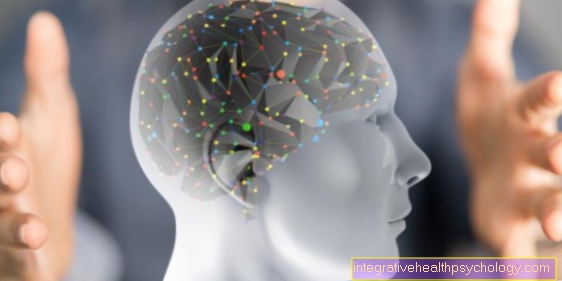
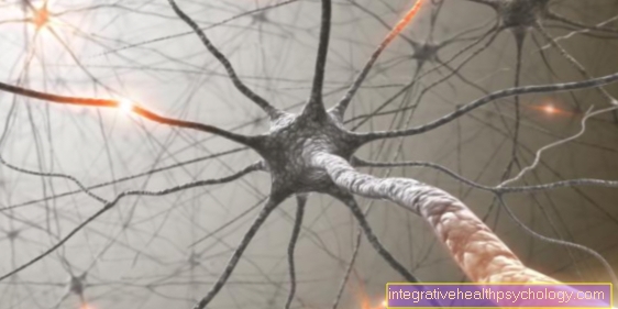
Brain atrophy
What is brain atrophy?
Brain atrophy is also known colloquially as brain atrophy. These terms are used to describe the loss of brain tissue due to age or disease.
This means that if the loss of mass and volume of the brain due to the death of nerve cells exceeds the age-related norm, then there is brain atrophy.
It is often associated with various neurological diseases. A distinction is made between generalized and focal brain atrophy.
Generalized brain atrophy affects all areas of the brain. In focal brain atrophy, only certain areas are affected.
Depending on the extent and location of the shrinkage, different failure symptoms and disorders can occur.

causes
Brain atrophy can have a variety of causes. It can be triggered by acute and chronic diseases.
Acute triggers can be traumatic brain injuries and severe strokes. In these cases, the acute irreversible death of nerve cells occurs, which can lead to brain atrophy.
The chronic causal diseases include, for example, multiple sclerosis, certain epilepsies, syphilis, AIDS and dementias such as Alzheimer's disease. In addition, drug abuse, certain long-term medications and alcoholism can lead to brain atrophy.
In addition, various eating disorders due to malnutrition can cause brain wasting.
According to some authors, severe or recurring depression can also lead to the death of nerve cells and thus to a loss of mass and volume in the brain.
What role does alcohol play?
Various studies have found that regular alcohol consumption can lead to brain atrophy.
Here experts speak of the rule of 3:
- about 1/3 of those affected do not develop brain atrophy,
- 1/3 results in reversible cerebral atrophy
- and another third has irreversible brain atrophy.
Some authors suggest a relationship between the amino acid homocysteine and alcohol-related brain atrophy.
The toxic amino acid homocysteine is produced in the body when the amino acid methionine is broken down. An increased concentration of homocysteine in the blood damages the blood vessels. This can affect the heart and the brain. If the brain is no longer supplied with sufficient blood, it can lead to brain atrophy. In addition, homcysteine occupies certain receptors in the brain and thus prevents physiological regulation. Eventually, this leads to nerve cells dying.
There are various factors and diseases that can lead to increased homocysteine levels. Regular alcohol consumption can also increase the level of homocysteine in the blood. Since folic acid is the natural antagonist, it can partly put the toxic effects of homocysteine into perspective.
The damage in the brain particularly affects the hippocampus and parts of the frontal lobe.
Various studies have found that brain atrophy can be reversible again after abstinence. This means that it is theoretically possible that more than 100,000 nerve cells can be formed each month. However, this is not possible with extreme and prolonged consumption of alcohol. In addition, it should be noted that permanent alcohol consumption promotes unforeseeable temporary and permanent, serious damage and diseases of various organs.
Causes of Frontal Brain Atrophy?
Frontal brain atrophy can have several causes.
The frontal area corresponds to the region of the brain, which is located behind the forehead. Acute events in the area, such as a pronounced stroke or severe traumatic brain injury, can lead to frontal brain atrophy.
In addition, chronic diseases such as Pick's disease can cause a loss of brain tissue in the forehead and temples. This condition is also known as frontotemporal dementia.
There are also subtypes of Alzheimer's disease that are located in the frontal area and can trigger brain atrophy there.
You might also be interested in our next article: : Traumatic brain injury
Brain atrophy in multiple sclerosis?
Various studies have shown that brain atrophy can develop in the context of multiple sclerosis and that this causes certain symptoms.
Some authors estimate the brain wasting per year in a person with multiple sclerosis to be about the size of a tablespoon.
This estimate applies to people with moderate disease. This information does not match with very light and very difficult forms.
In the meantime, new treatment options can reduce brain wasting to around a teaspoon.
For a long time it was thought that the symptoms and relapses of the disease were due to the damaged myelin sheaths. However, it has now been established that brain atrophy can damage the gray matter of the brain. An increased breakdown of the nerve cell processes and the synapses causes an intensification and progression of the symptoms. Therefore, in addition to avoiding relapses, reducing brain wasting is an essential treatment goal in multiple sclerosis.
Since brain atrophy happens slowly, it is often only noticed when it is already irreversible. Regular check-ups are therefore essential if you have multiple sclerosis.
The next article may also be of interest to you: Therapy of Multiple Sclerosis
diagnosis
Depending on what caused the brain atrophy and whether it was acute or insidious, it will be recognized by the patient or relatives at an earlier or later point in time.
If the onset is gradual, a doctor is often only consulted late. This takes an own and a third-party anamnesis.
That means he asks the patient himself and also the relatives.
In the event of abnormalities, appropriate screening procedures are carried out to test cognitive, linguistic, neuropsychological and sensorimotor performance. That means orientation skills, various memory, concentration and attention skills, as well as visual-spatial perception, responsiveness, coordination, language motor skills and language understanding are tested. In addition, mood swings and lows, personality changes and drive are recorded.
If there are restrictions in one of the areas, these will be examined more closely. In addition, it is necessary to perform imaging procedures such as computed tomography (CT), magnetic resonance tomography (MRT) or positron emission tomography (PET).
You can read more information on this topic here:
- Radiation exposure in computed tomography
- Is an MRI harmful?
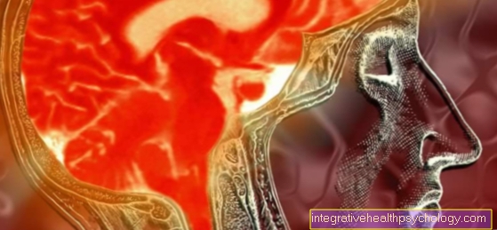
What can you see of the brain in the MRI?
Magnetic resonance imaging (MRI) can be used to visualize the different types of tissue in the brain.
Mathematical calculations by the computer create sectional images in which the various levels and layers of the brain are shown. In addition, tumors and inflammations can be displayed. However, bones are not easily recognizable. The unchanged images from the MRI are black and white.
Whether areas appear darker or lighter depends on the hydrogen content of the respective brain tissue. An even more differentiated image can be obtained by injecting a contrast medium. The contrast agent is lighter in color and enables blood vessels to be distinguished from the surrounding tissue.
In addition, the contrast agent often collects in tumors and enables them to be better represented and their exact location and size to be recorded.
Read more on the subject below: Procedure of an MRI
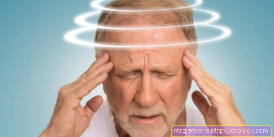
Symptoms - which symptoms suggest brain atrophy?
Depending on the affected brain area, the symptoms can be very different. Often a reduced drive and a change in personality with a loss of interest and social withdrawal can be the first signs.
In technical jargon, this symptom is also known as apathy.
In addition, psychological changes that manifest themselves in mood swings, affect lability, disinhibition and depression can indicate brain atrophy.
This harbors the risk that cerebral atrophy will be mistakenly “only” interpreted as depression and accordingly treated incorrectly.
In addition, cognitive limitations can occur that are often not noticed by the patient himself. Depending on the level of education and intelligence, these limitations can be compensated for for a long time.
The cognitive limitations can turn out to be disorders
- in memory,
- problem-solving behavior,
- orientation,
- the attention,
- concentration,
- spatial-visual perception,
- in thinking
- and show in action.
Furthermore, impaired speech production, motor impairments and also a restricted ability to smell can indicate brain atrophy.
With pronounced brain atrophy, seizures and hallucinations can occur.
Read more about this under: Drugs for Depression
Treatment and therapy
The therapy for brain atrophy depends on the causative disease. The goal of any treatment is to stop the progression of brain atrophy. Accordingly, attempts are made to treat the underlying disease adequately.
If the brain atrophy is due to drug or alcohol abuse, withdrawal therapy should be carried out. If the brain atrophy is caused by bacterial infection, antibiotics are used.
If viruses are responsible for the brain atrophy, antivirals are administered. In the case of dementia, multiple sclerosis and epilepsy, individual attempts are made to find a way to reduce the loss of brain mass.
The treatment can include medicamentous as well as occupational, physical, speech and neuropsychotherapy and neurosurgical measures.
Empathetic and competent advice and support from relatives is also very important.
You can read more information about the therapy of brain atrophy under: Occupational therapy
Course of a brain atrophy
Brain atrophy can occur suddenly as a result of an acute event such as a severe stroke and cause nerve cells to die off very quickly. In these cases, the brain atrophy does not have to progress in general.
If brain atrophy has arisen due to degenerative diseases, the onset is usually insidious. In these cases, the brain atrophy is progressive (progressive).
frequently asked Questions
Is Brain Atrophy Reversible?
Overall, brain atrophy is often irreversible. This means that the nerve cells that have died usually do not regenerate.
However, within certain limits it is possible that new nerve cells will form.
Depending on the cause of the brain atrophy, it can be stopped. This means that if you abstain from alcohol and drugs, for example, the brain atrophy no longer persists.
In degenerative diseases such as multiple sclerosis or Alzheimer's disease, the progression can only be delayed, but not stopped.
That is, the breakdown of brain tissue continues.
Is Brain Atrophy Hereditary?
Brain atrophy per se is not inherited, but a few underlying conditions that can cause brain atrophy can be inherited.
For example, Pick's disease is inherited.
How does brain atrophy affect life expectancy?
It used to be that brain atrophy involved minimizing life expectancy. In the meantime the medical possibilities have developed further, so that the assumption that a brain atrophy reduces the life expectancy cannot (any longer) be maintained.
In the meantime, there are more efficient examination options as well as more targeted medicinal, surgical and non-medicinal measures that can counteract brain atrophy to a certain extent or have compensatory benefits.
Accordingly, brain atrophy does not generally correlate with life expectancy. This means that there is generally no lower life expectancy in the case of brain atrophy.
Depending on which other symptoms, influences and conditions of the disease that triggered the brain atrophy are present, life expectancy can be differently high or low.
What is the relationship between brain atrophy and dementia?
In dementia, various pathological mechanisms lead to the death of nerve cells and thus to brain atrophy.
The cause of the respective form of dementia can be different.
So far, 4 possible causes have been identified.
These include circulatory problems, the presence of Lewy bodies, amyloid plaques, and deposits of a protein called tau.
The various forms of dementia differ in a few typical characteristics, but all of them cause disorders in thinking, acting, language and coordination.
Read more on the subject below: Circulatory disorders
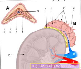
What is cerebellar atrophy?
With cerebellar atrophy, brain matter in the cerebellum disappears.
The causes are divided into three groups:
- the hereditary,
- the sporadic and
- the symptomatic form.
The hereditary forms are the cerebellar atrophies, which are inherited.
The symptomatic forms are triggered, for example, by drugs, viruses or alcohol. The sporadic forms are accepted when both other forms can be excluded.
The forms differ according to the symptoms and have characteristic features, but all have in common that the cerebellum atrophies. As a result, failures occur that affect the actual tasks of the cerebellum. This means that those affected often have problems with the coordination of movements in the body, impaired sensibility, impaired eye motor skills and cognitive deficits.
The four classic symptoms of cerebellar atrophy are ataxia, dysmetria, intention tremor, and dysarthria.
- Ataxia is characterized by uncoordinated, uncontrolled movements of the trunk, arms, and legs.
- Dysmetry describes grasping or aiming or pointing past a target.
- An intention tremor is a tremor of the arms, which shows itself when reaching for a goal.
- Dysarthria is a speech disorder that manifests itself in a choppy, slurred, or otherwise altered articulation.
Read more on the subject below: Cerebellar atrophy and cerebellar damage