How can you recognize tooth decay?
introduction
The initial stage of tooth decay often develops without symptoms, which is why the person concerned usually does not notice it. The patient only visits the dentist when the first pain occurs. In this case, however, the tooth is usually irreversibly damaged.
In general, the carious lesion is difficult to see visually, as it can vary greatly in color, shape and size. Caries can be yellowish, light brownish or black and the entry point of the disease can be so small that it can hardly be seen with the naked eye.
One of the most important criteria for the presence of tooth decay for the dentist is the softened, mushy appearing hard tooth substance, which differs significantly from the healthy hard tooth enamel.
This could also be of interest to you: Symptoms of tooth decay

For carious changes under a filling, crown or in the interdental space, it is impossible for the person concerned to recognize them himself. The dentist also needs aids for this, such as a probe or x-rays.
Therefore, it makes sense in any case to visit the treating dentist as soon as possible in the event of color changes or pain in the teeth in order to diagnose and treat caries or to rule out a disease.
You might also be interested in this topic: Molar Incisors Hypomineralization
Can you spot tooth decay yourself at home?
Due to the different dimensions and the variety of colors that a caries lesion can take on, it is difficult for a layperson to recognize it, so that it is often only noticed when there is pain that something is wrong. The patient himself should actively and regularly look into his mouth while brushing his teeth in order to detect changes as quickly as possible.
However, this can only be achieved to a limited extent, since the lighting conditions are usually not good and the teeth are always moist due to the flow of saliva. The dentist always assesses the teeth in a saliva-free, dry state in order to identify changes in color and structure in the teeth.
Furthermore, a change in color does not mean caries. Black discolorations, especially in the depths of the fissures, are most inactive forms of caries that can only be treated with regular fluoridation and show no tendency to spread.
When eating berries or drinking tea, coffee and cola or red wine, discoloration can quickly appear, which can be mistaken for tooth decay. If this discoloration affects many or even all teeth, it can usually be assumed that it is not caries.
In order for the patient to be sure, however, he should consult the dentist in the event of a suspected diagnosis, who can confirm or invalidate it. Regular checks once or twice a year are therefore sensible and recommended.
How can you find out caries in a milk tooth?
A caries in the deciduous dentition can spread much faster than with the permanent teeth due to the structural differences of the deciduous teeth. The thinned and softer enamel layer and the reduced mineral content make the milk tooth more prone to tooth decay.
In the early stages of this disease, white to yellowish, often punctiform indentations can be seen in the top layer of enamel. The tooth becomes rough in the affected area. Since parents are often unable to recognize these early stages of caries, the children usually come to the dentist too late and very advanced caries lesions develop quickly (as in the Nursing Bottle Syndrome).
By constantly drinking sweetened liquids through the feeding bottle, the front teeth are so badly damaged by caries that only black stubs of the teeth remain, which have to be extracted. Dental treatment is usually very stressful for small children, which is why there are specially trained children's dentists. Their aim is to use alternative stunning methods such as Laughing gas, to spare the child a lengthy and painful treatment, so that no dental phobia develops.
In general, parents should regularly look into the child's oral cavity and check their oral hygiene in order to identify changes as quickly as possible so that caries is not only noticed in an advanced state.
Read also under: Tooth decay in children - cause for concern?
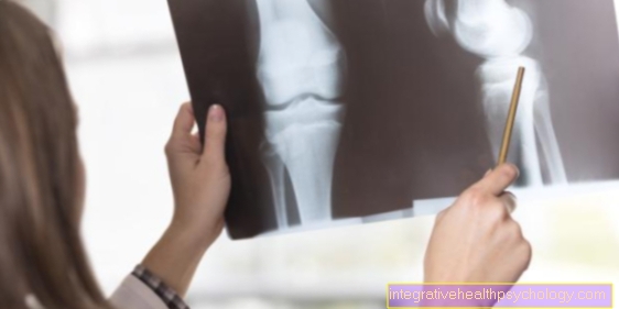
Detecting tooth decay in the X-ray
Caries can be diagnosed in the X-ray examination. The different densities and thicknesses of the tissue types influence the absorption capacity for X-rays. The enamel is denser than the dentin and appears whiter in the X-ray image. If the tooth now has a carious lesion, this changes the density of the hard tooth substance at this point, which makes it appear different in the X-ray image than the rest of the tooth.
Since the caries decomposes the tooth structure, the lesions appear dark to completely black in the X-ray film. The density is lower at this point than before. This is a good way of assessing how deep and large the tooth decay is and how it can be treated.
How can you diagnose tooth decay under a filling?
Caries under a filling cannot be detected by mere inspection. The only way to detect so-called secondary caries is x-ray diagnostics. In the majority of cases, bitewing diagnostics are used to identify caries between the teeth.
For those affected it is almost impossible to perceive tooth decay under a filling. It only becomes noticeable if the filling loosens due to the loosened bond due to caries or even completely detaches and falls off.
If there is caries under the filling, the adhesive layer with which the filling is attached will loosen as it progresses. As a result, the filling is no longer firmly connected to the tooth part. If the filling has fallen out, the defect is visible and the patient can see the caries. The area under the filling usually appears yellow, light brown or dark brown in color and can be painful and / or sensitive to cold.
In this case, it is best to make an appointment with the dentist immediately in order to treat the defect and remove the caries as quickly as possible.
See also under: Filling fell out - what to do?
How can you recognize tooth decay under a crown?
In most cases, tooth decay is invisible under a crown, as the crown covers the entire tooth.
Most of the time, no caries can be detected in an X-ray image either, since the crown material almost completely absorbs the X-rays and therefore nothing of the inside of the crown is reflected on the X-ray film.
Only when the caries reaches the portion under the crown would it be visible on the X-ray film.
For the dentist, the defect under the crown is only noticeable when it loosens or the crown edges no longer seal tightly. When touching the probe, it is possible to get under the crown margin, which is not allowed to happen with a sufficient supply.
Therapeutically, the crown must then be removed to treat the tooth decay under the crown. The tooth decay can be so deep that only a root canal treatment can save the tooth. In this case, a post build-up should also be made after the root filling to stabilize the weakened tooth. After the treatment, a new crown must be made.
Caries diagnosis in the interdental spaces
The interdental space is predisposed to the development of caries, as it is difficult to clean there and food residues are often overlooked. The main reason for this insidious, because it goes unnoticed, form of tooth decay is the insufficient use of dental floss or interdental brushes.
In the posterior region in particular, it is hardly possible to see it, so that the dentist usually cannot see anything either. For this reason, there are special X-ray images, so-called bitewing images, which only show the crown portion of the teeth and the spaces between the teeth. Other x-rays, such as the OPG or single-tooth films are not suitable for diagnosing caries between the teeth because they cause distortions and overlays.
When evaluating the bitewing image, it can often be observed that two adjacent teeth have a caries lesion that share a space between the teeth. This means that two teeth are carious and therefore two fillings are required for therapy.
For this reason, it is advisable to have these x-rays taken regularly during dental check-ups (about every two years or more often depending on the patient's risk of caries) in order to be able to assess the interdental spaces. The admission is usually a cash benefit.
Find out more at: X-ray for caries diagnosis


.jpg)