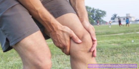
Illustration popliteal pain

Back of the knee pain
A - tear of the outer band / inner band
B - injuries to the menisci
C - osteoarthritis
D - popliteal cyst / Baker's cyst
E - thrombosis
- Inner meniscus -
Medial meniscus - Inner band -
Ligament collateral tibial - Popliteal muscle -
Popliteus muscle - Shin - Tibia
- Internal calf muscle -
M. gastrocnemius, caput mediale - External calf muscle -
M. gastrocnemius, caput laterale - Two-headed hamstrings
Biceps femoris muscle - Half-tendon muscle -
Semitendinosus muscle - Femur - Femur
- Posterior cruciate ligament -
Posterior cruciate ligament - Articular cartilage -
Cartilago articularis - Anterior cruciate ligament -
Ligament cruciatum anterius - Outer meniscus -
Lateral meniscus - Outer band -
Ligamentum collaterale fibulare - Fibula - Fibula
You can find an overview of all Dr-Gumpert images at: medical illustrations
Related images

Illustration
Baker's cyst

Illustration
posterior cruciate ligament

Illustration
Torn knee ligament

Illustration
Inner knee ligament tear

Illustration
Knee joint

Illustration
Kneecap

Illustration
Outside knee pain

Illustration
Inside knee pain

Illustration
Cartilage damage

Illustration
Cruciate ligament

Illustration
meniscus

Illustration
anterior cruciate ligament