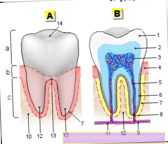
Figure tooth anatomy

a - tooth crown - Corona dentis
b - tooth neck - Cervix dentis
c - tooth root - Radix dentis
- Tooth enamel -
Enamelum - Dentin (= dentine) -
Dentinum - Tooth pulp in the tooth cavity -
Pulp dentis in Cavitas dentis - Gums -
Gingiva - Root canal
- Cement -
Cementum - Root skin - Periodontium
- Opening of the tooth root tip -
Foramen apicale dentis - Nerve fibers
- Alveolar bone (tooth-bearing
Part of the jawbone) -
Pars alveolaris
(Alveolar process) - Blood vessels
- Tooth root tip -
Apex denitis - Point of division of the tooth roots
(Fork) - Bifurcation - Tooth furrow
You can find an overview of all Dr-Gumpert images at: medical illustrations
Related images

Illustration
Inflammation of the gums

Illustration
Tooth scheme