Zygomatic bone
introduction
The cheekbone (cheekbones, cheekbones, lat. Os zygomaticum) is a pair of bones of the facial skull. It is located on the lateral edge of the eye sockets and is instrumental in the lateral contour of the face.
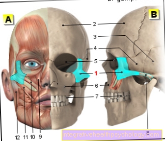
Illustration of the zygomatic bone

- Zygomatic bone -
Os zygomaticum - Frontal bone - Frontal bone
- Temporal bone - Temporal bone
- Sphenoid bone - Sphenoid bone
- Eye socket - orbit
- Upper jaw - Maxilla
- Molar tooth -
Dens moralis - Zygomatic arch -
Arcus zygomaticus - Upper lip lifter -
Levator muscle
labii superioris - Small zygomatic muscle -
Zygomaticus minor muscle - Zygomatic large muscle -
Zygomaticus major muscle - Masseter (jaw muscle) -
Muscle masseter
A - skull from the front
(Muscles and bones)
B - skull from the left
You can find an overview of all Dr-Gumpert images at: medical illustrations
topography
The zygomatic bone lies in front of the temporal bone (Temporal bone) and below the frontal bone (Frontal bone) and the sphenoid (Sphenoid bone).
It lies over the maxillary bone (Maxilla) and forms most of the lateral wall of the bony eye socket (Orbit).
Function of the zygomatic bone
The cheekbone directs the chewing pressurewhich of the great Molars goes out, now and then forms the wall of the eye and nasal cavities.
In addition, it articulates and is with the adjacent bony structures Point of origin of some facial muscles.
construction
The cheekbone points three faces on:
- a lateral (Lateral facies),
- a lying to the eye socket (Orbital facies) and
- a adjacent to the temporal bone (Facies temporalis) Surface.
The lateral face has a convex shape and contains one in its center Bone opening, as Zygomaticofacial foramen referred to as. In this area the large and small zygomatic muscles (Os zygomaticus major and minor) Their origin. Through the soft tissues of the face, this side can be called "Cheekbones" feel.
The side facing the temporal bone (Facies temporalis) shows one concave inner surface. It falls backwards (dorsally) and towards the middle (medial). In its upper part it forms one small pit, the Temporal fossa in the lower area the infratemporal fossa. In the front area of this surface there is a rough, almost triangular bone area, which is connected to the upper jaw bone (maxilla). Also within this area there is a small hole that Zygomaticotemporal foramen.
The side facing the eye socket (orbital face) is smooth and, together with the maxillary bone and the sphenoid bone, forms part of the wall and floor of the bony eye socket. The small one is roughly in the middle Zygomatic Orbitals foramen. The cheekbone shows a small one upwards bony process on the Frontal process.
This articulates with the zygomatic process of the frontal bone (frontal bone). A further bony process is the Maxillary process. It has a clumsy, triangular cross-section and articulates with the zygomatic process of the upper jawbone (maxilla). At its front edge lies the Origin of the levator labii superioris muscle.
It points backwards Temporal processwhich in turn is connected to the zygomatic process of the temporal bone (os temporalis). These two bony parts together form the zygomatic arch from (Arcus zygomaticus), at the lower edge of which the Origin of the large masticatory muscle (Masseter muscle) lies.
clinic
This can be done through strong violence Break the cheekbone and to severe pain to lead. this is a typical injury in boxers after being hit in the face. Therapy is either conservative, or but operational with the help of a plate.
Zygomatic fracture
A cheekbone fracture is one Fracture of the cheekbone, mostly through external force. Since neighboring facial bones are also often affected, it becomes the lateral midface fractures summarized.
This group is further subdivided according to the location and severity of the break. It is also important whether the fragments are shifted against one another, i.e. whether they are dislocated.
Overall, the lateral midface fracture makes over 50% of all midface bone bridges out. The fault lines often follow the Adhesion lines of the individual skull bones, the so-called Sutures.
The symptoms of a zygomatic fracture are next to one painful swelling the affected side often one Change in the geometry of the face. Due to the origin of the chewing muscles on the zygomatic arch, chewing is usually very strong painful and only possible to a limited extent. If the eye socket is involved, you can Double vision occur.
Therapy is used for dislocated bones operational carried out. Here the bones are realigned. This allows them to grow together again and usually heal without consequences.
Cheekbone contusion
A cheekbone contusion is caused by strong force on the cheekbone. She can do a lot painful be and expresses itself through a severe swelling on the affected side of the face.
A cooling is therefore very helpful for treatment. It relieves both pain and swelling. Pain relieving ointments can also be used, but should be applied with caution due to their proximity to the eye. Since the cheekbone contusion is very similar to the symptoms Zygomatic fracture shows you should see a doctor.
This is especially true with one very severe swellingthat too eye affects, gets stronger and does not resolve within a few days. With additional symptoms such as Double vision, severe headache, nausea or Vomit a doctor should be consulted immediately.
Same goes for Faint as a result of the violence. When in doubt, a X-ray image be made to the The suspicion of a cheekbone fracture can be safely excluded. If this is not the case, no further treatment is required.
Summary
The cheekbone is a small bony portion of the facial skullwhich is connected to the adjacent bony structures via smaller processes. He makes you large part of the bony eye socket (Orbita) and is also significantly on the lateral facial contour involved.

